

A BOLD search for baseline
- PMID: 17223362
- PMCID: PMC2684871
- DOI: 10.1016/j.neuroimage.2006.11.035
A BOLD search for baseline
Abstract
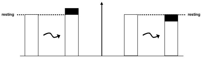
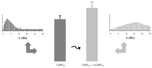
While we occasionally observe negative BOLD signals, its physiological basis has remained uncertain. This is in part due to the qualitative use of fMRI where the baseline is conveniently differenced away to reveal focal area(s) of interest. Recently, however, there has been a noticeable trend towards quantitative neuroimaging where changes in oxidative energetics (CMR(O2)) are quantified by calibrated fMRI. Pasley et al. [Pasley, B.N., Inglis, B.A., Freeman, R.D., 2007. Analysis of oxygen metabolism implies a neural origin for the negative BOLD response in human visual cortex. NeuroImage] used calibrated fMRI in conjunction with a novel stimulus paradigm to investigate the neural basis of the negative BOLD signal in awake humans. They hypothesized - based on prior results - that if the baseline was lowered then DeltaCMR(O2) would have to be larger. While their main findings point to an energetic basis for the negative BOLD signal, their results have far reaching implications for the present definition of baseline as well as for future research investigating the neural and/or energetic basis of baseline.
Figures
References
-
- Angel A, Berridge DA, Unwin J. The effect of anaesthetic agents on primary cortical evoked responses. Br J Anaesth. 1973;45:824–836. - PubMed
-
- Armstrong-James M, Fox K. Spatiotemporal convergence and divergence in the rat S1 “barrel” cortex. J Comp Neurol. 1987;263:265–281. - PubMed
-
- Chapin JK. Laminar differences in sizes, shapes, and response profiles of cutaneous receptive fields in the rat SI cortex. Exp Brain Res. 1986;62:549–559. - PubMed
-
- Chapin JK, Waterhouse BD, Woodward DJ. Differences in cutaneous sensory response properties of single somatosensory cortical neurons in awake and halothane anesthetized rats. Brain Res Bull. 1981;6:63–70. - PubMed
Publication types
MeSH terms
Substances
Grants and funding
LinkOut - more resources
Full Text Sources
Medical
