Review
. 2007 Jan;20(1):79-114.
doi: 10.1128/CMR.00015-06.

Modes and modulations of antibiotic resistance gene expression

Affiliations
Review

Modes and modulations of antibiotic resistance gene expression

Florence Depardieu et al. Clin Microbiol Rev. 2007 Jan.

Abstract

Since antibiotic resistance usually affords a gain of function, there is an associated biological cost resulting in a loss of fitness of the bacterial host. Considering that antibiotic resistance is most often only transiently advantageous to bacteria, an efficient and elegant way for them to escape the lethal action of drugs is the alteration of resistance gene expression. It appears that expression of bacterial resistance to antibiotics is frequently regulated, which indicates that modulation of gene expression probably reflects a good compromise between energy saving and adjustment to a rapidly evolving environment. Modulation of gene expression can occur at the transcriptional or translational level following mutations or the movement of mobile genetic elements and may involve induction by the antibiotic. In the latter case, the antibiotic can have a triple activity: as an antibacterial agent, as an inducer of resistance to itself, and as an inducer of the dissemination of resistance determinants. We will review certain mechanisms, all reversible, that bacteria have elaborated to achieve antibiotic resistance by the fine-tuning of the expression of genetic information.


Figures

FIG. 1.
FIG. 1.
Schematicrepresentation of a two-component regulatory system. Structural features of sensor (top) and regulator (bottom) proteins. H, N, G1, F, and G2 refer to the motifs conserved in histidine protein kinases and are shown as hatched blue boxes. The phosphorylated histidine is nested in a highly conserved sequence termed the H box, close to the N-terminal border of the conserved kinase domain. The G1 and G2 domains are glycine rich and resemble nucleotide binding motifs seen in other proteins. The sequences of the remaining D and F boxes reveal little about their possible functions. In the regulator, the central aspartate is the site of phosphorylation, whereas the amino-terminal pair is probably important for catalysis. The conserved lysine may be involved in effecting the phosphorylation-induced conformational changes that regulate output activity. Asp, aspartate; His, histidine; P, phosphate; dotted blue box, sensor domain; blue box, transmembrane domain; white box, kinase domain; horizontally striped green box, receiver domain; checkerboard green box, effector domain. a.a., amino acids.
FIG. 2.
FIG. 2.
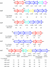
Comparison of the van gene clusters. Open arrows represent coding sequences (red arrows, regulatory genes; purple arrows, genes required for resistance; blue arrows, accessory genes; pink and yellow arrows, genes of unknown function) and indicate the direction of transcription. The percentages of amino acid (aa) identity between the deduced proteins of reference strains BM4147 (VanA) (19), V583 (VanB) (77), BM4339 (VanD) (42), BM4174 (VanC) (10), BM4405 (VanE) (1), and BM4518 (VanG) (63) are indicated under the arrows. The vertical bar in vanYG indicates the frameshift mutation leading to a predicted truncated protein. NA, not applicable.
FIG. 3.
FIG. 3.
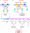
Model for positive (phosphorylation) and negative (dephosphorylation) control of VanR by VanS and schematic representation of the synthesis of peptidoglycan precursors in VanA- or VanB-type strains. Kinase (A) and phosphatase (B) activities of VanS are depicted. K, heterologous kinase; R, regulator; S, sensor. Dotted blue circle, sensor domain; blue box, transmembrane domain; white circle, kinase domain; horizontally striped green circle, receiver domain; checkerboard green box, effector domain.
FIG. 4.
FIG. 4.
(A) Schematic representation of the binding of VanR-type regulators to the vanA and vanB promoters and (B) comparison of affinity of VanR or VanRB and VanR-P or VanRB-P for DNA fragments carrying the PR/PRB and PH/PYB promoters. Open arrows represent coding sequences (red arrows, regulatory genes; purple arrows, genes required for resistance; black arrows, genes of unknown function).
FIG. 5.
FIG. 5.
Schematic representation of the VanSB sensor and location of amino acid substitutions in teicoplanin-resistant mutants. H, N, G1, F, and G2 refer to the motifs conserved in histidine protein kinases and are shown as hatched boxes. The putative membrane-associated sensor domain (dotted blue) containing transmembrane segments (blue) and the putative cytoplasmic kinase domain (white) are indicated. Het, heterogeneously resistant; R, resistant; S, sensitive; Te, teicoplanin; Vm, vancomycin.
FIG. 6.
FIG. 6.
Schematic representation of the cell membranes with examples of multidrug efflux systems. ABC, ATP binding cassette; MFP, membrane fusion protein; MFS, major facilitator superfamily; OM, outer membrane; OMF, outer membrane factor; RND, resistance nodulation cell division; SMR, small multidrug resistance.
FIG. 7.
FIG. 7.
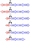
Genetic organization of the adeRS-adeABC operon from A. baumannii, the smeRS-smeABC operon from S. maltophilia, and the mexR-mexAB-oprM, mexT-mexEF-oprN, nfxB-mexCD-oprJ, and mexZ-mexXY MDR operons from P. aeruginosa. Purple arrows, structural genes for drug efflux complexes; red arrows, regulatory genes that either repress (−) or activate (+) gene expression (this still has to be confirmed for mexZ).
FIG. 8.
FIG. 8.
Characteristics of IS elements. DR, direct repeat; IR, inverted repeat; −35/−10 and −35, approximate locations of promoter consensus sequences.
FIG. 9.
FIG. 9.
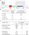
Transcriptional control in class 1 integrons. (A) Schematic representation of the integron platform. 5′ CS and 3′ CS, 5′- and 3′-conserved segments, respectively; Pc and P2, promoter regions (see the text); attI1, recombination site; C1 and C2, gene cassettes; 59 be, 59-base-pair elements with possible stem-loop structures. (B) Relative strength of integron-borne promoter variants. aDetermined relative to the strength of the tac promoter, set at 1 (data are from reference 141). bStreptomycin concentration at which 50% of cells plated formed colonies (data are from reference 54). (C) Effects of cassette order on resistance levels. aResistance is conferred to streptomycin (Sm) by aadA2, to gentamicin (Gm) by aacC1, and to kanamycin (Km) by aacA4 (the genes for which a position effect is observed and the corresponding antibiotic concentrations at which 50% of cells plated formed colonies [IC50s] are shown in boldface type). bIC50 data are from reference .
FIG. 10.
FIG. 10.
Possible functions of ORF-11 in the translation of cassette-associated genes. SD, Shine-Dalgarno sequence. (A) Translational coupling (a, b, and c) (see the text). The position of the stop codon in the recombination core site is underlined. (B) Fusion. (C) Unknown effect of ORF-11.
FIG. 11.
FIG. 11.
Relative ORF-11-mediated translation efficiencies. C, control; WT, configuration depicted in Fig. 10A (wild type); Δa, Δb, and Δc, mutants with deletion of fragments a, b, and c as shown in Fig. 10A (data are from reference 98).
FIG. 12.
FIG. 12.
Alternative conformations of the mRNA from the inducible erm(C) gene of pE194. Shown is the secondary structure of the mRNA in the absence (A) or presence (B) of erythromycin. RBS, ribosome binding site; LP, leader peptide; ORF, open reading frame; 1, 2, 3, and 4, inverted repeat. Green and red lines indicate the coding sequence.
FIG. 13.
FIG. 13.
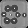
S. aureus containing an erm(C) gene that is inducibly expressed. CM, clindamycin; E, erythromycin; L, lincomycin; SP, spiramycin; PI, pristinamycin IA (streptogramin factor B); PII, pristinamycin IIA (streptogramin factor A); PT, pristinamycin; TEL, telithromycin. A D-shaped zone can be observed for the clindamycin (and noninducer macrolides) zone of inhibition on the edge closest to the erythromycin zone of inhibition.
FIG. 14.
FIG. 14.
S. aureus containing an erm(C) gene that is constitutively expressed. CM, clindamycin; E, erythromycin; L, lincomycin; SP, spiramycin; PI, pristinamycin IA (streptogramin factor B); PII, pristinamycin IIA (streptogramin factor A); PT, pristinamycin; TEL, telithromycin.

Similar articles

Cited by

References

    1. Abadía, L., P. Courvalin, and B. Périchon. 2002. vanE gene cluster of vancomycin-resistant Enterococcus faecalis BM4405. J. Bacteriol. 184:6457-6464. - PMC - PubMed
    1. Abadia-Patino, L., K. Christiansen, J. Bell, P. Courvalin, and B. Perichon. 2004. VanE-type vancomycin-resistant Enterococcus faecalis clinical isolates from Australia. Antimicrob. Agents Chemother. 48:4882-4885. - PMC - PubMed
    1. Abo-Amer, A. E., J. Munn, K. Jackson, M. Aktas, P. Golby, D. J. Kelly, and S. C. Andrews. 2004. DNA interaction and phosphotransfer of the C4-dicarboxylate-responsive DcuS-DcuR two-component regulatory system from Escherichia coli. J. Bacteriol. 186:1879-1889. - PMC - PubMed
    1. Aendekerk, S., B. Ghysels, P. Cornelis, and C. Baysse. 2002. Characterization of a new efflux pump, MexGHI-OpmD, from Pseudomonas aeruginosa that confers resistance to vanadium. Microbiology 148:2371-2381. - PubMed
    1. Aires, J. R., T. Kohler, H. Nikaido, and P. Plesiat. 1999. Involvement of an active efflux system in the natural resistance of Pseudomonas aeruginosa to aminoglycosides. Antimicrob. Agents Chemother. 43:2624-2628. - PMC - PubMed

MeSH terms

Substances