

A mobile tryptophan is the intrinsic charge transfer donor in a flavoenzyme essential for nikkomycin antibiotic biosynthesis
- PMID: 17223703
- PMCID: PMC2716209
- DOI: 10.1021/bi062087s
A mobile tryptophan is the intrinsic charge transfer donor in a flavoenzyme essential for nikkomycin antibiotic biosynthesis
Abstract
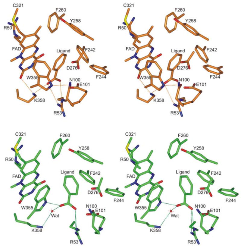
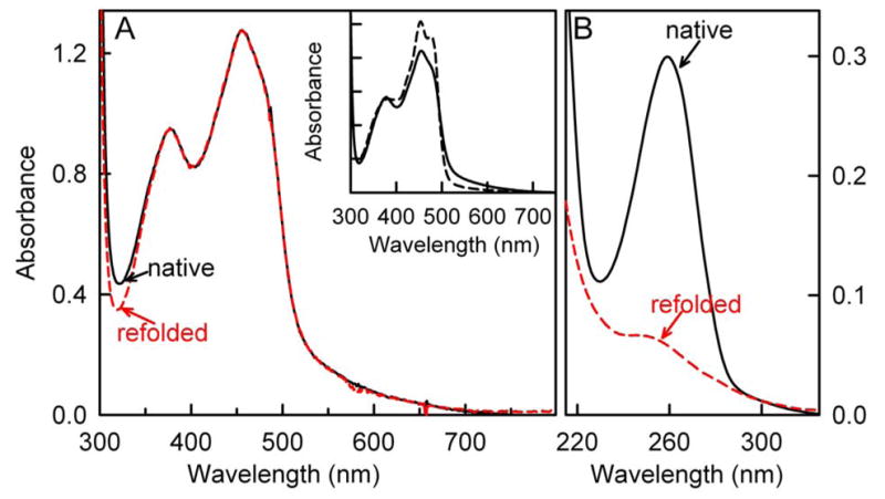
The flavoenzyme nikD is required for the biosynthesis of nikkomycin antibiotics. NikD exhibits an unusual long wavelength absorption band attributed to a charge transfer complex of FAD with an unknown charge transfer donor. NikD crystals contain an endogenous active site ligand. At least four different compounds are detected in nikD extracts, including variable amounts of two ADP derivatives that bind to the enzyme's dinucleotide binding motif in competition with FAD, picolinate (0.07 mol/mol of nikD) and an unknown picolinate-like compound. Picolinate, the product of the physiological catalytic reaction, matches the properties deduced for the active site ligand in nikD crystals. The charge transfer band is eliminated upon mixing nikD with excess picolinate but not by a reversible unfolding procedure that removes the picolinate-like compound, ruling out both compounds as the intrinsic charge transfer donor. Mutation of Trp355 to Phe eliminates the charge transfer band, accompanied by a 30-fold decrease in substrate binding affinity. The results provide definitive evidence for Trp355 as the intrinsic charge transfer donor. The indole ring of Trp355 is coplanar with or perpendicular to the flavin ring in "open" or "closed" crystalline forms of nikD, respectively. Importantly, a coplanar configuration is required for charge transfer interaction. Absorption in the long wavelength region therefore constitutes a valuable probe for monitoring conformational changes in solution that are likely to be important in nikD catalysis.
Figures


References
-
- Fiedler HP, Kurth R, Langharig J, Delzer J, Zahner H. Nikkomycins: Microbial inhibitors of chitin synthetase. J Chem Biotechnol. 1982;32:271–280.
-
- Bruntner C, Bormann C. The Streptomyces tendae Tu901 L-lysine 2-aminotransferase catalyzes the initial reaction in nikkomycin D biosynthesis. Eur J Biochem. 1998;254:347–355. - PubMed
-
- Bruntner C, Lauer B, Schwarz W, Mohrle V, Bormann C. Molecular characterization of co-transcribed genes from Streptomyces tendae Tu901 involved in the biosynthesis of the peptidyl moiety of the peptidyl nucleoside antibiotic nikkomycin. Mol Gen Genet. 1999;262:102–114. - PubMed
-
- Venci D, Zhao G, Jorns MS. Molecular characterization of nikD, a new flavoenzyme important in the biosynthesis of nikkomycin antibiotics. Biochemistry. 2002;41:15795–15802. - PubMed
Publication types
MeSH terms
Substances
Grants and funding
LinkOut - more resources
Full Text Sources
