. 2007 May 1;403(3):409-20.
doi: 10.1042/BJ20061364.

Pseudomonas aeruginosa and sPLA2 IB stimulate ABCA1-mediated phospholipid efflux via ERK-activation of PPARalpha-RXR

Affiliations

Pseudomonas aeruginosa and sPLA2 IB stimulate ABCA1-mediated phospholipid efflux via ERK-activation of PPARalpha-RXR

Marianna Agassandian et al. Biochem J. .

Abstract

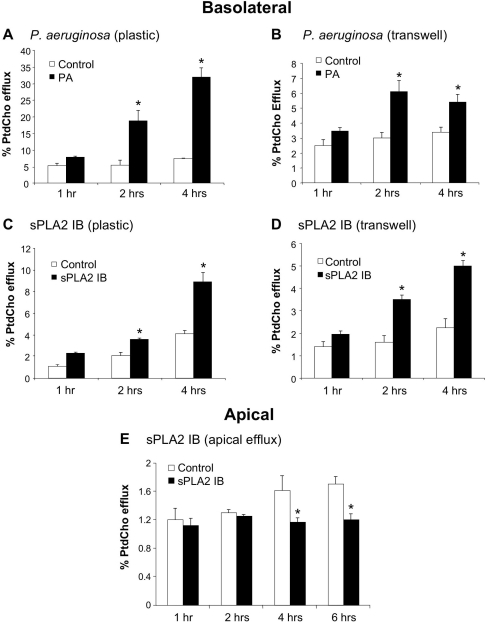
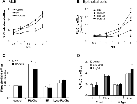
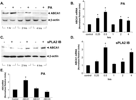
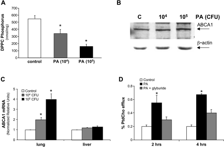
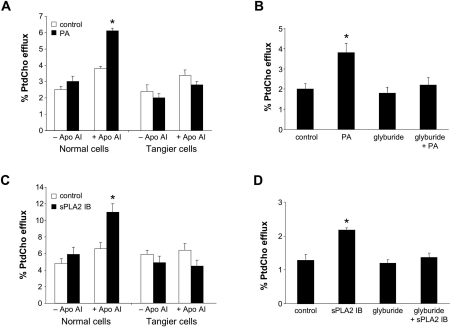
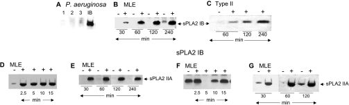
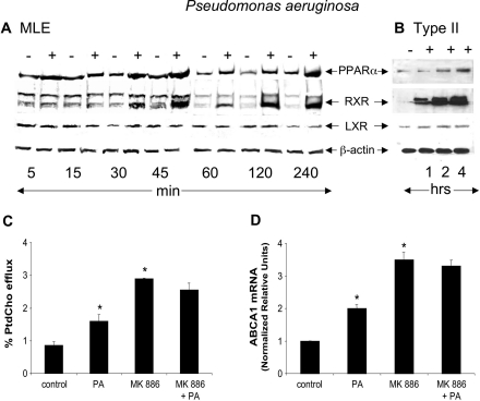
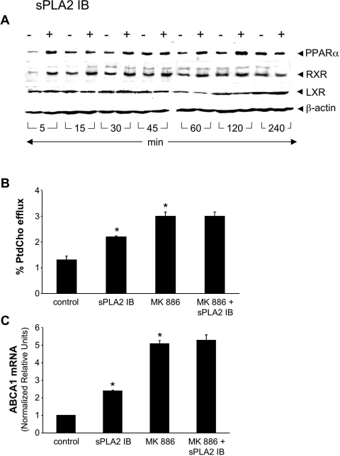
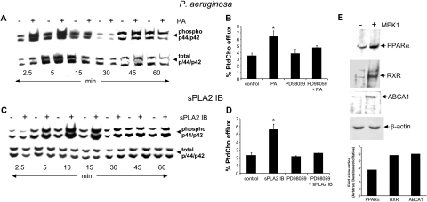
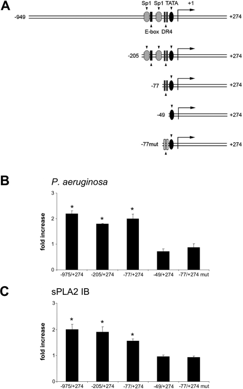
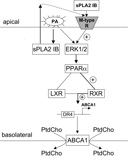
Bacterial infection triggers an acute inflammatory response that might alter phospholipid metabolism. We have investigated the acute-phase response of murine lung epithelia to Pseudomonas aeruginosa infection. Ps. aeruginosa triggered secretion of the pro-inflammatory lipase, sPLA2 IB (phospholipase A2 IB), from lung epithelium. Ps. aeruginosa and sPLA2 IB each stimulated basolateral PtdCho (phosphatidylcholine) efflux in lung epithelial cells. Pre-treatment of cells with glyburide, an inhibitor of the lipid-export pump, ABCA1 (ATP-binding cassette transporter A1), attenuated Ps. aeruginosa and sPLA2 IB stimulation of PtdCho efflux. Effects of Ps. aeruginosa and sPLA2 IB were completely abolished in human Tangier disease fibroblasts, cells that harbour an ABCA1 genetic defect. Ps. aeruginosa and sPLA2 IB induced the heterodimeric receptors, PPARa (peroxisome-proliferator-activated receptor-a) and RXR (retinoid X receptor), factors known to modulate ABCA1 gene expression. Ps. aeruginosa and sPLA2 IB stimulation of PtdCho efflux was blocked with PD98059, a p44/42 kinase inhibitor. Transfection with MEK1 (mitogen-activated protein kinase/extracellular-signal-regulated kinase kinase 1), a kinase upstream of p44/42, increased PPARa and RXR expression co-ordinately with increased ABCA1 protein. These results suggest that pro-inflammatory effects of Ps. aeruginosa involve release of an sPLA2 of epithelial origin that, in part, via distinct signalling molecules, transactivates the ABCA1 gene, leading to export of phospholipid.


Figures

Figure 1
Figure 1. Ps. aeruginosa and sPLA2 IB induce PtdCho efflux in lung epithelia
Effect of Ps. aeruginosa (PA) on basolateral PtdCho efflux was assayed in MLE-12 cells plated in 60 mm plastic and transwell dishes, labelled with [methyl-3H]choline and in the presence of 20 μg/ml HDL as a PtdCho acceptor. Cells were exposed to Ps. aeruginosa (MOI=5) for various times (A, B). Effect of 100 μg/ml sPLA2 IB on basolateral (C, D) and apical (E) efflux was measured under similar conditions. The cells and basolateral and apical medium were harvested separately after exposure to Ps. aeruginosa and sPLA2 IB and processed for PtdCho analysis. *P<0.05 compared with control. Results are means±S.E.M. for three to five independent experiments.
Figure 2
Figure 2. Ps. aeruginosa triggers phospholipid efflux selectively
(A) Effect of Ps. aeruginosa (PA) and sPLA2 IB on cholesterol efflux in MLE-12 cells plated in transwells and labelled with [3H]cholesterol. Cells were exposed to Ps. aeruginosa (MOI=5) and sPLA2 IB (100 μg/ml) for various times after labelling and processed for cholesterol efflux. (B) Effect of Ps. aeruginosa on PtdCho efflux in different types of epithelial cells. (C) Effect of Ps. aeruginosa (PA) and sPLA2 IB on efflux of SM and lyso-PtdCho in MLE-12 cells after 2 h of exposure to agonists. (D) Effect of LPS from E. coli and Salmonella Typhi (S. Typhi) on PtdCho efflux in MLE-12 cells. *P<0.05 compared with control. Results are means±S.E.M. for three independent experiments.
Figure 3
Figure 3. Ps. aeruginosa and sPLA2 IB stimulate expression of ABCA1 in lung epithelial cells
MLE-12 cells were incubated with (+) or without (−) Ps. aeruginosa (MOI=5) (PA) (A, B) or sPLA2 (100 μg/ml) (C, D) for various times. (A, C) ABCA1 and β-actin levels were determined by immunoblotting using ∼100 μg of cell lysate protein, resolution by SDS/10% PAGE and probing with anti-ABCA1 or β-actin polyclonal antibodies. (B, D) Ps. aeruginosa and sPLA2 induce ABCA1 mRNA levels in MLE-12 cells. Cultured cells were infected with or without Ps. aeruginosa for various times or cultured with sPLA2, and total cellular RNA was harvested for analysis of ABCA1 mRNA by real-time PCR. (E) MLE-12 cells were exposed to Ps. aeruginosa (MOI=5) (PA) for 2 h, and total cellular RNA was harvested for analysis of ABCG1 and ABCG5 mRNA by real-time PCR. Results are mean±S.E.M. relative units, which were normalized to murine GAPDH, for three independent experiments. *P<0.05 compared with control.
Figure 4
Figure 4. Ps. aeruginosa regulates PtdCho export in vivo
C57BL/6J mice were intratracheally administrated with Ps. aeruginosa strain PA103 (PA) using either control agar particles or bacterial agar particles (104–105 cfu). After 1 h, mice were killed, the lungs were lavaged, and surfactant pellets were isolated. (A) Surfactant pellets were analysed for DPPC levels, expressed as nmol of lipid phosphorus/mg of protein. (B) The lungs were processed for ABCA1 and β-actin levels by immunoblot analysis. (C) Total mRNA was harvested from lung and liver for analysis of ABCA1 mRNA by real-time PCR. (D) Primary mouse type II cells were isolated and labelled with [methyl-3H]choline for 20 h, exposed to glyburide (250 μM) for 1 h, and then incubated with or without PA103 (MOI=5) in the presence of HDL (20 μg/ml). Medium and cells were harvested for PtdCho analysis. *P<0.05 compared with control. Results are means±S.E.M. for three independent experiments.
Figure 5
Figure 5. Ps. aeruginosa and sPLA2 IB regulate PtdCho export via ABCA1 in lung epithelia
(A, C) Immortalized HSFs obtained from normal and Tangier disease patients were plated on plastic dishes, labelled with [methyl-3H]choline for 20 h, and then incubated with or without Ps. aeruginosa (MOI=5) (PA) (A) or sPLA IB (100 μg/ml) (C) using apoAI (10 μg/ml) as a PtdCho acceptor. Medium and cells were harvested after 2 h of exposure to Ps. aeruginosa or sPLA2 IB and processed for PtdCho analysis. (B, D) Effect of pharmacological ABCA1 inhibition on Ps. aeruginosa (B) or sPLA2 IB (D) -induced basolateral efflux was assayed in transwell dishes. Cells were labelled with [methyl-3H]choline for 20 h, exposed to glyburide (250 μM) and then incubated with or without Ps. aeruginosa (MOI=5) or sPLA2 IB (100 μg/ml) for 2 h in the presence of HDL (20 μg/ml). Medium and cells were harvested for PtdCho analysis. *P<0.05 compared with control. Results are means±S.E.M. for three to four independent experiments.
Figure 6
Figure 6. Ps. aeruginosa and sPLA2 IB stimulate secretion of sPLA IIA and sPLA2 IB in lung epithelia
(A) Ps. aeruginosa was incubated in serum-free Hite's medium without antibiotics and in the absence of MLE-12 cells for 2 h. Proteins from 5 ml of the medium were precipitated with 60% TCA and 1.5% sodium desoxycholate and applied on SDS/PAGE at various concentrations: lane 1, 50 μg; lane 2, 100 μg; lane 3, 150 μg. Gels were then processed for sPLA2 IB immunoblotting. In lane 4, bovine pancreas sPLA2 IB (5 μg) was loaded on to the gel as a positive control. MLE-12 (B, DG) and mouse primary type II cells (C) were incubated with (+) or without (−) Ps. aeruginosa (MOI=5) (BE) or sPLA2 IB (100 μg/ml) (F, G) for various times, and medium and cell lysates were harvested. Proteins from 5 ml of the medium was precipitated with 60% TCA and 1.5% sodium desoxycholate, and immunoblot analysis was performed with anti-(sPLA2 IB) and anti-(sPLA2 IIA) polyclonal antibodies. Bovine pancreas sPLA2 (5 μg) was loaded on the gel as a positive control. Immunoblots are representative of three independent experiments.
Figure 7
Figure 7. Ps. aeruginosa up-regulates PPARα and RXR in MLE-12 cells
MLE-12 (A) and primary mouse type II (B) cells were incubated with (+) or without (−) Ps. aeruginosa (MOI=5) for various times. Cell lysates were harvested, separated by SDS/10% PAGE, and probed with anti-ABCA1, anti-PPARα, anti-RXR, anti-LXR and anti-β-actin antibodies. Relative protein levels in each experiment were detected by stripping one nitrocellulose membrane and re-probing with various antibodies. (C) Basolateral PtdCho efflux. MLE-12 cells were plated on transwells, labelled with [methyl-3H]choline for 20 h, incubated with MK 886 (5 μM) for 1 h, and then infected with Ps. aeruginosa (MOI=5) for 2 h using HDL (20 μg/ml) as a PtdCho acceptor. Basolateral medium and cells were harvested for PtdCho analysis. (D) MLE-12 cells were incubated with MK 886 and Ps. aeruginosa as described above, and total RNA was harvested for ABCA1 mRNA analysis by real-time PCR using GAPDH transcripts as an internal control. *P<0.05 compared with control. Results in histograms are means±S.E.M. for three independent experiments.
Figure 8
Figure 8. sPLA2 IB up-regulates PPARα and RXR in MLE-12 cells
(A) MLE-12 cells were incubated with (+) or without (−) sPLA2IB (100 μg/ml) for various times. Cell lysates were harvested, separated by SDS/10% PAGE, and probed with anti-ABCA1, anti-PPARα, anti-RXR, anti-LXR and anti-β-actin antibodies. Relative protein levels in each experiment were detected by stripping one nitrocellulose membrane and reprobing with various antibodies. (B) Basolateral PtdCho efflux. MLE-12 cells were plated on transwells, labelled with [methyl-3H]choline for 20 h, incubated with MK 886 (5 μM) for 1 h and then exposed to sPLA2 IB (100 μg/ml) for 2 h using HDL (20 μg/ml) as a PtdCho acceptor. Basolateral medium and cells were harvested for PtdCho analysis. (C) MLE-12 cells were incubated with MK 886 and sPLA2 IB as described above, and total RNA was harvested for ABCA1 mRNA analysis by real-time PCR using GAPDH transcripts as an internal control. *P<0.05 compared with control. Results in histograms are means±S.E.M. for three independent experiments.
Figure 9
Figure 9. Ps. aeruginosa and sPLA2 IB activate p44/p42 kinase
MLE-12 cells were exposed to Ps. aeruginosa (MOI=5) (PA) (A) or sPLA2 IB (100 μg/ml) (C) for various times, and cell lysates were harvested and processed for immunoblotting. Nitrocellulose membranes were probed with antibodies against phosphorylated (active) and total p44/p42 isoforms. (B) Basolateral PtdCho export. MLE-12 cells were labelled for 20 h with [methyl-3H]choline, incubated for 1 h with PD98059 (10 μM) and subsequently exposed for 2 h to Ps. aeruginosa (B) or sPLA2 IB (D). Medium was harvested and processed for PtdCho analysis. (E) MLE-12 cells were transfected with a MEK1 plasmid (4 μg) overnight, and the next day harvested and processed for immunoblot analysis using anti-PPARα, anti-RXR, anti-ABCA1 and anti-β-actin antibodies. *P<0.05 compared with control. Results in histograms are means±S.E.M. for three independent experiments.
Figure 10
Figure 10. Ps. aeruginosa and sPLA2 IB increase ABCA1 promoter activity
(A) Schematic illustration of ABCA1 promoter constructs. Three constructs contain a DR-4 element (−949/+274, −205/+274 and −77/+274), one lacks this element (−49/+274), and one contains a mutation within DR4 (−77/+274 mut). (B, C) Cells were co-transfected with these ABCA1 promoter reporters and pSV-β-galactosidase for 2–4 h before exposure to Ps. aeruginosa (MOI=5) (B) or PLA2 IB (100 μg/ml) (C) for 2 h. Lysates were assayed for luciferase and β-galactosidase activities. Results are means±S.E.M. fold increases of luciferase/β-galactosidase activities in the cells exposed to Ps. aeruginosa and sPLA2 IB compared with control for three independent experiments. *P<0.05 compared with control.
Figure 11
Figure 11. Mechanisms of ABCA1 activation by Ps. aeruginosa and exogenous sPLA2 IB
The solid lines indicate known pathways, the broken lines indicate putative mechanisms, and the thick solid lines indicate mechanisms discussed in the present paper.

Similar articles

Cited by

References

    1. Sadikot R. T., Blackwell T. S., Christman J. W., Prince A. S. Pathogen–host interactions in Pseudomonas aeruginosa pneumonia. Am. J. Respir. Crit. Care Med. 2005;171:1209–1223. - PMC - PubMed
    1. Hauser A. R., Cobb E., Bodi M., Mariscal D., Valles J., Engel J. N., Rello J. Type III protein secretion is associated with poor clinical outcomes in patients with ventilator-associated pneumonia caused by Pseudomonas aeruginosa. Crit. Care Med. 2002;30:521–528. - PubMed
    1. Rabin S. D., Hauser A. R. Pseudomonas aeruginosa ExoU, a toxin transported by the type III secretion system, kills Saccharomyces cerevisiae. Infect. Immun. 2003;71:4144–4150. - PMC - PubMed
    1. Arbibe L., Vial D., Rosinski-Chupin I., Havet N., Huerre M., Vargaftig B. B., Touqui L. Endotoxin induces expression of type II phospholipase A2 in macrophages during acute lung injury in guinea pigs: involvement of TNF-α in lipopolysaccharide-induced type II phospholipase A2 synthesis. J. Immunol. 1997;159:391–400. - PubMed
    1. Vial D., Senorale-Pose M., Havet N., Molio L., Vargaftig B. B., Touqui L. Expression of the type-II phospholipase A2 in alveolar macrophages: down-regulation by an inflammatory signal. J. Biol. Chem. 1995;270:17327–17332. - PubMed

Publication types

MeSH terms