. 2007 Feb;147(2):330-8.
doi: 10.1111/j.1365-2249.2006.03284.x.

Immunological responses and protective immunity against tuberculosis conferred by vaccination of Balb/C mice with the attenuated Mycobacterium tuberculosis (phoP) SO2 strain

Affiliations

Immunological responses and protective immunity against tuberculosis conferred by vaccination of Balb/C mice with the attenuated Mycobacterium tuberculosis (phoP) SO2 strain

D Aguilar et al. Clin Exp Immunol. 2007 Feb.

Abstract

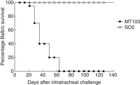
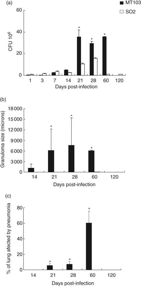
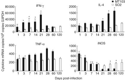
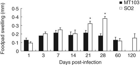
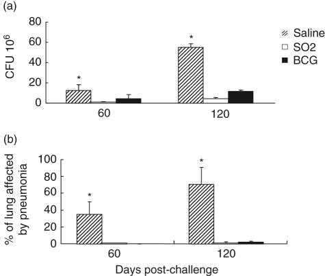
The Mycobacterium tuberculosis phoP mutant strain SO2 has been shown previously to be more attenuated than Mycobacterium bovis bacillus Calmette-Guérin (BCG) and confers protective immunity against tuberculosis in mice and guinea pig models. In this study we have investigated the survival and immunological responses of Balb/c mice infected with the M. tuberculosis SO2 strain. All Balb/C mice survived intratracheal infection with M. tuberculosis SO2 strain under conditions where all the mice infected with the parental M. tuberculosis MT103 had died after 9 weeks. Infection of Balb/c mice with M. tuberculosis SO2 was associated with comparatively lower levels of interferon (IFN)-gamma, interleukin (IL)-4 and tumour necrosis factor (TNF)-alpha and higher levels of inducible nitric oxide synthase (iNOS) during the late stage of infection, when compared with M. tuberculosis MT103 infection. The delayed-type hypersensitivity (DTH) response against M. tuberculosis culture filtrates was similar in mice infected with either the M. tuberculosis phoP SO2 strain or M. tuberculosis MT103. The protective efficacy of M. tuberculosis SO2 was compared with M. bovis BCG when delivered subcutaneously to groups of Balb/C mice. Following intratracheal challenge with M. tuberculosis H37Rv, protection was generated by 60 days post-challenge in mice vaccinated with either vaccine. At day 120 post-challenge the levels of protection were still significantly greater when compared with the non-vaccinated control group. The levels of protection conferred by vaccination with M. tuberculosis SO2 or with M. bovis BCG were similar, as measured by granuloma coalescence and pneumonia in addition to growth reduction of M. tuberculosis H37Rv.


Figures

Fig. 1
Fig. 1
Attenuation of Mycobacterium tuberculosis SO2 in Balb/c mice: survival of Balb/c mice infected with M. tuberculosis MT103 or the attenuated M. tuberculosis SO2 monitored over 120 days post-infection.
Fig. 2
Fig. 2
Lung bacillary burden and histomorphometry of Balb/c mice infected with Mycobacterium tuberculosis SO2 or MT103 parental strain by the intratracheal route. (a) Numbers of colony-forming units (CFUs) in the lungs of mice infected with either M. tuberculosis MT103 or M. tuberculosis SO2; (b) granuloma size (μ2) in M. tuberculosis MT103 or M. tuberculosis SO2-infected lungs; (c) percentage of lung surface area affected by pneumonia. Bars represent means ± s.d. from three mice per time-point. Asterisks represent statistical significance between groups at those time-points (P < 0·05).
Fig. 3
Fig. 3
Representative histology and mycobacterial DNA (IS6110) detection by in situ polymerase chain reaction (PCR) in mice lung after intratracheal infection with Mycobacterium tuberculosis SO2. (a) Occasional and small granulomas (arrows) are seen after 60 days post-infection; (b) at 120 days post-infection, middle hyperplasia of lymphoid tissue associated with the bronchial wall (arrow) is the only distinctive histological abnormality; (c) large mediastinal lymph node with diffuse hyperplasia is generated after 120 days post-infection; (d) at 120 days post-infection, in situ PCR detection of mycobacterial DNA is positive (blue dots) in bronchial hyperplastic lymphoid tissue (arrows) and bronchial epithelium (arrow heads); (e) after 120 days of infection, mycobacterial DNA detection by in situ PCR also shows strong positivity in the venular endothelium (arrows) and bronchial epithelium (arrowheads); (f) positive PCR signal in macrophages located in the subcapsular sinus of mediastinal hyperplastic lymph node.
Fig. 4
Fig. 4
Immunological responses of Balb/C mice infected with Mycobacterium tuberculosis MT103 and M. tuberculosis SO2: mean ± s.d. values of quantitative expression of mRNA encoding cytokines and nitric oxide synthase (iNOS) determined by real-time polymerase chain reaction (PCR) in lung homogenates from three mice per group infected with either M. tuberculosis MT103 or M. tuberculosis SO2. Asterisks represent statistical significance between groups at those time-points (P < 0·05).
Fig. 5
Fig. 5
Delayed-type hypersensitivity response of Mycobacterium tuberculosis MT103 and M. tuberculosis SO2 in Balb/c mice: delayed-type hypersensitivity response measured in mouse footpads following injection of mycobacterial antigens. Bars represent means ± s.d. from three mice per time-point in two different experiments. Asterisks represent statistical significance between groups at those time-points (P < 0·05).
Fig. 6
Fig. 6
Protective efficacy of Mycobacterium tuberculosis SO2 and BCG-Phipps in Balb/c mice: (a) numbers of colony-forming units (CFUs) of M. tuberculosis H37Rv recovered from lungs of challenged mice that were vaccinated with either M. tuberculosis SO2 or M. bovis bacille Calmette–Guérin (BCG), compared with saline-treated controls; (b) percentage of pneumonia in Balb/c mice vaccinated subcutaneously with either M. tuberculosis SO2 or M. bovis BCG at 60 and 120 days following intratracheal challenge with M. tuberculosis H37Rv. Asterisks denotes where results from saline-treated mice controls are significantly different (P < 0·05) from mice receiving either vaccine.

References

    1. World Health Organization (WHO) Global Report tuberculosis. Geneva: WHO; 2005. Global tuberculosis control - surveillance, planning, financing.
    1. Young D, Dye C. The development and impact of tuberculosis vaccines. Cell. 2006;124:683–7. - PubMed
    1. Fine PE. Variation in protection by BCG: implications of and for heterologous immunity. Lancet. 1995;346:1339–45. - PubMed
    1. Hernandez-Pando R, Orozco H, Sampieri A, et al. Correlation between the kinetics of Th1, Th2 cells and pathology in a murine model of experimental pulmonary tuberculosis. Immunology. 1996;89:26–33. - PMC - PubMed
    1. Behr MA. BCG − different strains, different vaccines? Lancet Infect Dis. 2002;2:86–92. - PubMed

Publication types

MeSH terms