. 2007 Jan 15:2:1.
doi: 10.1186/1750-1326-2-1.

Angiotensin type 1 receptor antagonist losartan, reduces MPTP-induced degeneration of dopaminergic neurons in substantia nigra

Affiliations

Angiotensin type 1 receptor antagonist losartan, reduces MPTP-induced degeneration of dopaminergic neurons in substantia nigra

Tom N Grammatopoulos et al. Mol Neurodegener. .

Abstract

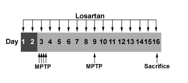
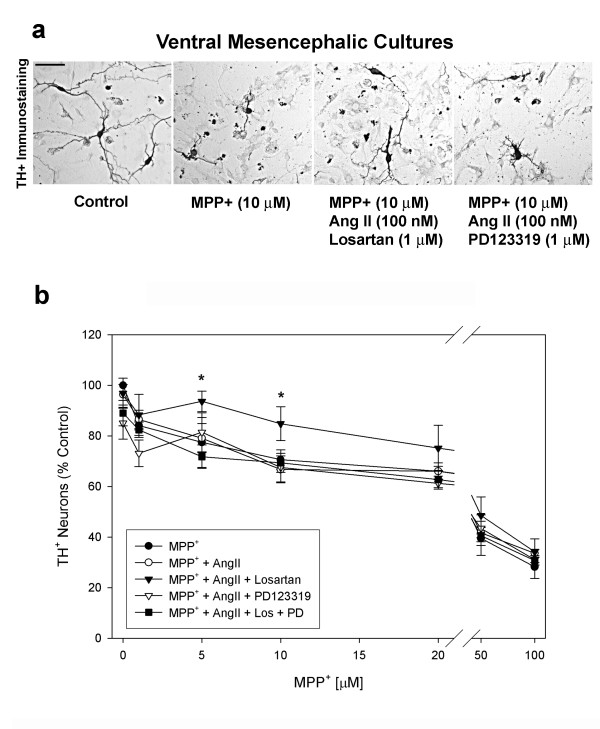
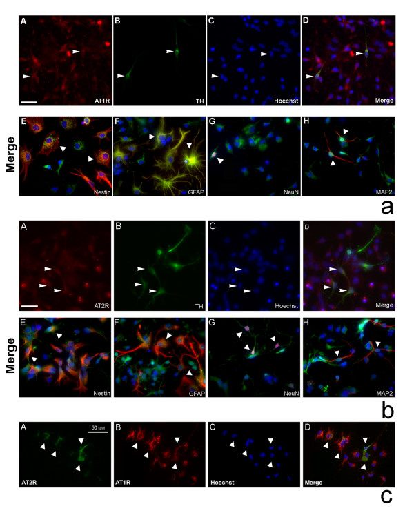
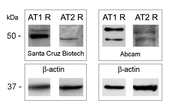
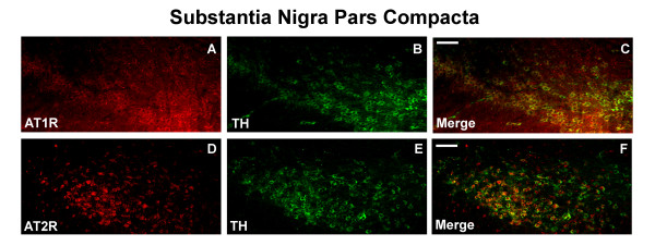
Background: Recent attention has focused on understanding the role of the brain-renin-angiotensin-system (RAS) in stroke and neurodegenerative diseases. Direct evidence of a role for the brain-RAS in Parkinson's disease (PD) comes from studies demonstrating the neuroprotective effect of RAS inhibitors in several neurotoxin based PD models. In this study, we show that an antagonist of the angiotensin II (Ang II) type 1 (AT1) receptor, losartan, protects dopaminergic (DA) neurons against 1-methyl-4-phenyl-1,2,3,6-tetrahydropyridine (MPTP) toxicity both in primary ventral mesencephalic (VM) cultures as well as in the substantia nigra pars compacta (SNpc) of C57BL/6 mice (Fig. 1).

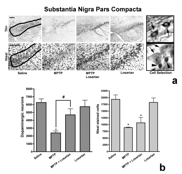
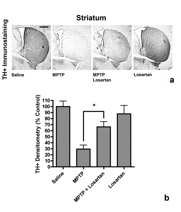
Results: In the presence of exogenous Ang II, losartan reduced MPP+ (5 muM) induced DA neuronal loss by 72% in vitro. Mice challenged with MPTP showed a 62% reduction in the number of DA neurons in the SNpc and a 71% decrease in tyrosine hydroxylase (TH) immunostaining of the striatum, whereas daily treatment with losartan lessened MPTP-induced loss of DA neurons to 25% and reduced the decrease in striatal TH+ immunostaining to 34% of control.

Conclusion: Our study demonstrates that the brain-RAS plays an important neuroprotective role in the MPTP model of PD and points to AT1 receptor as a potential novel target for neuroprotection.


Figures

Figure 1
Figure 1
Timeline of the experimental design. Arrows point to when subcutaneous injections of losartan (90 mg/kg) and intraperitoneal injections of MPTP-hydrochloride (20 mg/kg) were administered and when the animals were sacrificed.
Figure 2
Figure 2
Angiotensin II protects DA neurons in vitro from MPP+ toxicity only in the presence of the AT1 receptor antagonist losartan. (a) Microscopic view of TH+ (DA) neurons in VM cultures in the presence of media or MPP+ (10 μM) alone, MPP+ with Ang II (100 nM), and MPP+ with either losartan (1 μM) or PD123319 (1 μM). Scale bar equal to 50 μm. (b) Quantification of TH+ neuron counts in VM cultures treated with increasing concentrations of MPP+ (1–100 μM) in the presence or absence of Ang II (100 nM) and AT receptor antagonists, losartan (1 μM) (AT1R) and/or PD123319 (1 μM) (AT2R). Results are mean ± SEM. (n = 4–6). (*) Represents a significant difference (p ≤ 0.05, One-way ANOVA followed by Newman-Keuls post-hoc test) from MPP+.
Figure 3
Figure 3
In vitro AT receptor expression profiles in primary rat VM cultures. (a) VM cultures were stained for TH (DA neurons) (green) (A), AT1 receptor (red) (B), and counter stained with Hoechst dye (blue nuclear stain) (C). Merged image of A-C (D). Panels (E-H) are merged images of AT1 receptor (green) co-labeling with either nestin (neural progenitors; red) (E), GFAP (astrocytes; red) (F), NeuN (neurons; red) (G), or MAP2 (neuronal marker; red) (H), counter stained with Hoechst dye (blue nuclear stain) and visualized under fluorescence microscopy (×400). Arrowheads indicate examples of cells of various phenotypes that contain AT1 receptors. Scale bar equal to 50 μm. (b) VM cultures were stained for TH (green) (A), AT2 receptor (red) (B), and counter stained with Hoechst dye (C). Merged image of A-C (D). Panels (E-H) are merged images of AT2 receptor (green) double stained with either nestin (red) (E), GFAP (red) (F), NeuN (red) (G), or MAP2 (red) (H), counter stained with Hoechst dye and visualized under fluorescence microscopy. Arrowheads point to various cell types positive for the AT2 receptor (×400). Scale bar equals 50 μm. (c) Panels (A-D) show co-expression of AT1 (red) (B) and AT2 (green) (A) receptors counter stained with Hoechst dye (C). Merged image of A-C (D). Arrowheads show examples of cells co-expressing both types of AT receptors.
Figure 4
Figure 4
Western immunoblot detection of the AT receptors in N27 dopaminergic cell line using either the Abcam or the Santa Cruz antibodies. The AT1 receptor is detected as two proteins representing the AT1A (43 kDa) and AT1B (53 kDa) encoded by two different genes. The AT2 is detected at 43 kDa.
Figure 5
Figure 5
In vivo AT receptor profile of DA neurons in the substantia nigra pars compacta (SNpc) of adult C57BL/6 male mice. Coronal sections (40 μm) were stained for (A) AT1 receptor (red) or (D) AT2 receptor (red), (B, E) TH (DA neurons) (green) and (C, F) represent merged images. Scale bar equals to 200 μm.
Figure 6
Figure 6
Detection of Agtr2, TH and GADH mRNAs by real time RT-PCR in TH+ neurons of the SNpc and VTA. Relative concentrations were determined using a standard curve of known concentrations of whole brain cDNAs.
Figure 7
Figure 7
Losartan protects DA neurons of the SNpc from MPTP toxicity. (a) TH immunoreactive neurons and Nissl stained cells in the SNpc from mice treated with saline, MPTP, MPTP + losartan or losartan alone. Scale bars equal to 100 μm. Saline panel shows a representative contour selection of the A9 region. The Cell Selection panel shows an example of counted TH+ neurons (indicated with arrows) or Nissl-stained cells with large nuclei (indicated with arrows) and excluded Nissl-stained cells with small nuclei (indicated with arrowheads). (b) Quantification of the number of TH+ neurons and Nissl stained cells in the SNpc, revealed a significant decrease in the number TH+ neurons in MPTP treated mice when compared to saline control treated animals. MPTP-injected mice treated daily with losartan significantly decreased the MPTP-induced loss of TH+ neurons in the SNpc. Significance is indicated by (#) when compared to MPTP-alone injected mice and (*) when compared to saline-alone injected mice (p ≤ 0.05, One-way ANOVA followed by Newman-Keuls post-hoc test). Data are represented as mean ± SEM, n = 4–5 for TH immunostain and n = 3 for Nissl stain.
Figure 8
Figure 8
Losartan prevents MPTP-induced denervation of the striatum. (a) Coronal sections of TH immunoreactivity in the striatum of mice treated with saline, MPTP, MPTP + losartan or losartan alone. Scale bars equal to 500 μm. (b) Quantification of striatal densities, showing a significant decrease in the percent of TH+ immunoreactivity in MPTP-treated mice. MPTP-injected mice treated daily with losartan had significantly higher levels of TH+ immunoreactivity in the striatum when compared to MPTP alone-injected mice (*, p ≤ 0.05, One-way ANOVA followed by Newman-Keuls post-hoc test). Data are represented as mean ± SEM, (n = 4–5).

References

    1. Parkinson J. In: An essay on the shaking palsy. Neely, Jones, editor. London: printed by Whittingham and Rowland for Sherwood; 1817.
    1. Hornykiewicz O. Dopamine (3-hydroxytyramine) and brain function. Pharmacol Rev. 1966;18:925–64. - PubMed
    1. Dauer W, Przedborski S. Parkinson's disease: mechanisms and models. Neuron. 2003;39:889–909. doi: 10.1016/S0896-6273(03)00568-3. - DOI - PubMed
    1. Allam MF, Castillo AS, Navajas RS. Parkinson's disease risk factors: genetic, environmental, or both? Neurol Res. 2005;27:206–8. doi: 10.1179/016164105X22057. - DOI - PubMed
    1. Twelves D, Perkins KS, Counsell C. Systematic review of incidence studies of Parkinson's disease. Mov Disord. 2003;18:19–31. doi: 10.1002/mds.10305. - DOI - PubMed