. 2007 Jun 14;26(28):4059-70.
doi: 10.1038/sj.onc.1210188. Epub 2007 Jan 15.

Ubiquitin-independent degradation of p53 mediated by high-risk human papillomavirus protein E6

Affiliations

Ubiquitin-independent degradation of p53 mediated by high-risk human papillomavirus protein E6

S Camus et al. Oncogene. .

Abstract

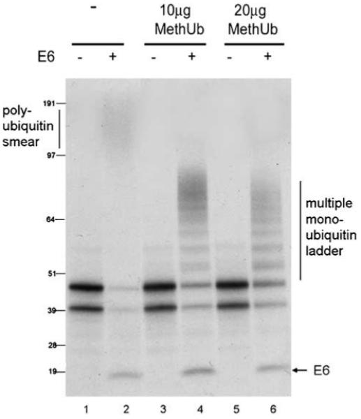
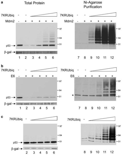
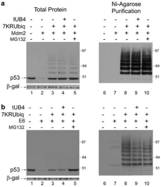
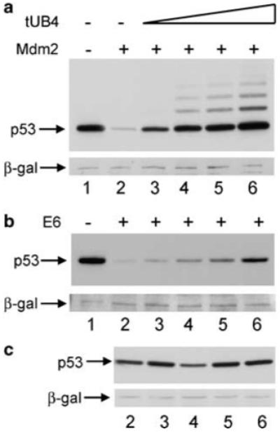
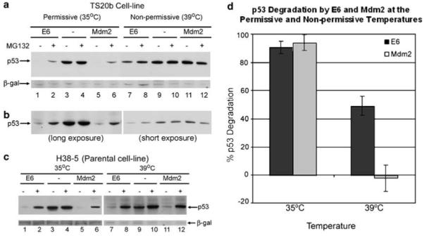
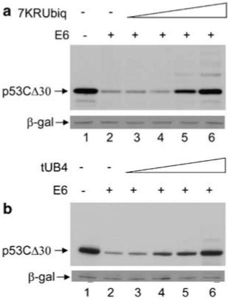
In vitro, high-risk human papillomavirus E6 proteins have been shown, in conjunction with E6-associated protein (E6AP), to mediate ubiquitination of p53 and its degradation by the 26S proteasome by a pathway that is thought to be analogous to Mdm2-mediated p53 degradation. However, differences in the requirements of E6/E6AP and Mdm2 to promote the degradation of p53, both in vivo and in vitro, suggest that these two E3 ligases may promote p53 degradation by distinct pathways. Using tools that disrupt ubiquitination and degradation, clear differences between E6- and Mdm2-mediated p53 degradation are presented. The consistent failure to fully protect p53 protein from E6-mediated degradation by disrupting the ubiquitin-degradation pathway provides the first evidence of an E6-dependent, ubiquitin-independent, p53 degradation pathway in vivo.


Figures

Figure 1
Figure 1
E6 mediates ubiquitination of p53 in vitro. p53, E6 or empty vector control (all under a T7 promoter) were translated separately in rabbit reticulocyte lysate in the presence of [35S]methionine. Translated p53 and pcDNA3 (lanes 1, 3 and 5) or E6 (lanes 2, 4 and 6) were mixed in the absence (lanes 1 and 2) or presence of 10 μg (lanes 3 and 4) or 20 μg (lanes 5and 6) purified methylated ubiquitin (MethUb) and further incubated at 25°C for 90 min.
Figure 2
Figure 2
7KR-ubiquitin only partially protects p53 from E6-mediated degradation. (a) H1299 cells were transfected with 1 μg p53 together with 2 μg Mdm2 (lanes 2-6 and 8-12) or control pcDNA3 vector (lanes 1 and 7). Co-expression of a titration of 5 μg (lanes 3 and 9), 10 μg (lanes 4 and 10), 15 μg (lanes 5 and 11) or 20 μg (lanes 6 and 12) His6-tagged 7KR-ubiquitin was performed. An Ni-agarose purification of His6-tagged 7KR-ubiquitinated species is shown in the right panel. (b) As in (a) but with E6 instead of Mdm2. (c) As in (a) (minus lanes 1 and 7) but with pcDNA3 instead of Mdm2. DNA concentrations are maintained with empty pcDNA3 control vector and β-gal is used as a loading and transfection-efficiency control throughout. p53 is detected with DO-1 antibody.
Figure 3
Figure 3
Effects of 7KR-ubiquitin and tUB4 on E6- and Mdm2-mediated degradation of p53. (a) H1299 cells were transfected with 0.5 μg p53 together with 1 μg Mdm2 (lanes 2-6), or control pcDNA3 vector (lane 1), as above. Twenty micrograms 7KR-ubiquitin was co-expressed in lane 3 and 20 μg 7KR-ubiquitin plus 20 μg tUB4 was co-expressed in the absence (lane 4) or presence (lane 5) of MG132, incubated for 3 h before harvesting. DNA concentrations were maintained with empty pcDNA3 control vector. (b) As in (a) but with E6 instead of Mdm2.
Figure 4
Figure 4
Tandem chains of four ubiquitin moieties only partially protect p53 from E6-mediated degradation. (a) H1299 cells were transfected with 0.5 μg p53 together with 1 μg Mdm2 (lanes 2-6) or control pcDNA3 vector (lane 1), as above. Co-expression of a titration of 5 μg (lane 3), 10 μg (lane 4), 15 μg (lane 5) or 20 μg (lane 6) pcDNA3 tUB4 was performed. (b) As in (a) but with E6 instead of Mdm2. (c), As in (a) (minus lane 1) but with pcDNA3 instead of Mdm2.
Figure 5
Figure 5
The non-canonical pathway of E6-mediated p53 degradation occurs at physiologically relevant levels of p53 and E6 expression. (a) H1299 cells were transfected with 0.1 μg p53 together with 0.2 μg E6 (lanes 2 and 4), Mdm2 (lanes 3 and 5) or control pcDNA3 vector (lane 1). (20 μg) tUB4 was co-transfected with E6 (lane 4) or Mdm2 (lane 5) as indicated. (b) H1299 cells transfected with 0.1 μg p53 in the absence (lane 2) or presence of 0.2 μg E6 (lane 3) or Mdm2 (lane 4) and samples were run beside an extract of the same total protein concentration of HeLa cell lysate (lane 1).
Figure 6
Figure 6
E6-mediated p53 degradation is only partially inhibited by inactivation of E1. (a) TS20b cells were transfected with p53 together with Mdm2 (lanes 5, 6, 11 and 12), E6 (lanes 1, 2, 7 and 8) or pcDNA3 (lanes 3, 4, 9and 10) and left at 35°C (lanes 1-6) or moved to the non-permissive temperature of 39°C (lanes 7-12) 20 h before harvesting. Cells were incubated for 3 h before harvesting with 20 μM MG132 as indicated (lanes 2, 4, 6, 8, 10 and 12). (b) As above but a longer exposure of the 35°C samples (lanes 1-6) and a shorter exposure of the 39°C samples (lanes 7-12). (c) Parental H38-5cells were transfected as in (a) and moved to 39°C 20 h before harvesting. (d) Graph showing degradation of p53 mediated by E6 (dark shaded bars) and Mdm2 (light shaded bars) at the permissive temperature (35°C) and the non-permissive temperature (39°C).
Figure 7
Figure 7
The C-terminus of p53 is important for ubiquitin independent degradation. (a) H1299 cells were transfected with 0.5 μg p53CΔ30 together with 1 μg E6 (lanes 2-6), or control pcDNA3 vector (lane 1). Co-expression of a titration of 5 μg (lane 3), 10 μg (lane 4), 15 μg (lane 5) or 20 μg (lane 6) pcDNA3 7KR-ubiquitin was performed with DNA concentrations maintained with empty pcDNA3 control vector. (b) As in (a) but with tUB4 instead of 7KR-ubiquitin.

References

    1. Benaroudj N, Tarcsa E, Cascio P, Goldberg AL. The unfolding of substrates and ubiquitin-independent protein degradation by proteasomes. Biochimie. 2001;83:311–318. - PubMed
    1. Bercovich Z, Kahana C. Involvement of the 20S proteasome in the degradation of ornithine decarboxylase. Eur J Biochem. 1993;213:205–210. - PubMed
    1. Camus S, Higgins M, Lane DP, Lain S. Differences in the ubiquitination of p53 by Mdm2 and the HPV protein E6. FEBS Lett. 2003;536:220–224. - PubMed
    1. Chen X, Chi Y, Bloecher A, Aebersold R, Clurman BE, Roberts JM. N-acetylation and ubiquitin-independent proteasomal degradation of p21(Cip1) Mol Cell. 2004;16:839–847. - PubMed
    1. Chowdary DR, Dermody JJ, Jha KK, Ozer HL. Accumulation of p53 in a mutant cell line defective in the ubiquitin pathway. Mol Cell Biol. 1994;14:1997–2003. - PMC - PubMed

Publication types