Review
. 2007 Feb;5(2):128-40.
doi: 10.1038/nrmicro1599.

Translational control of retroviruses

Affiliations
Review

Translational control of retroviruses

Laurent Balvay et al. Nat Rev Microbiol. 2007 Feb.

Abstract

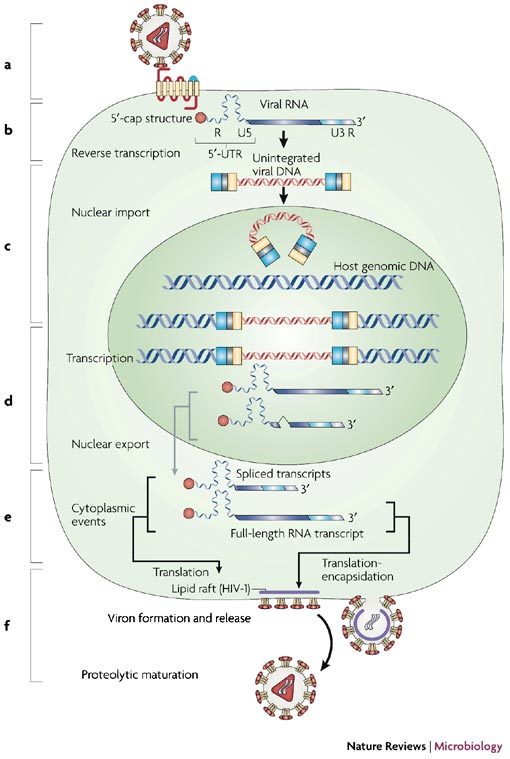
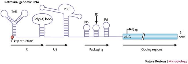
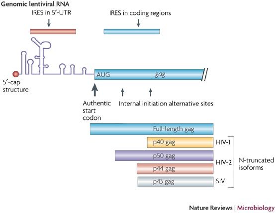
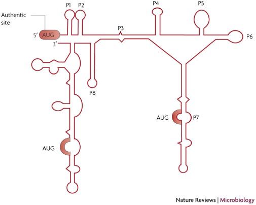
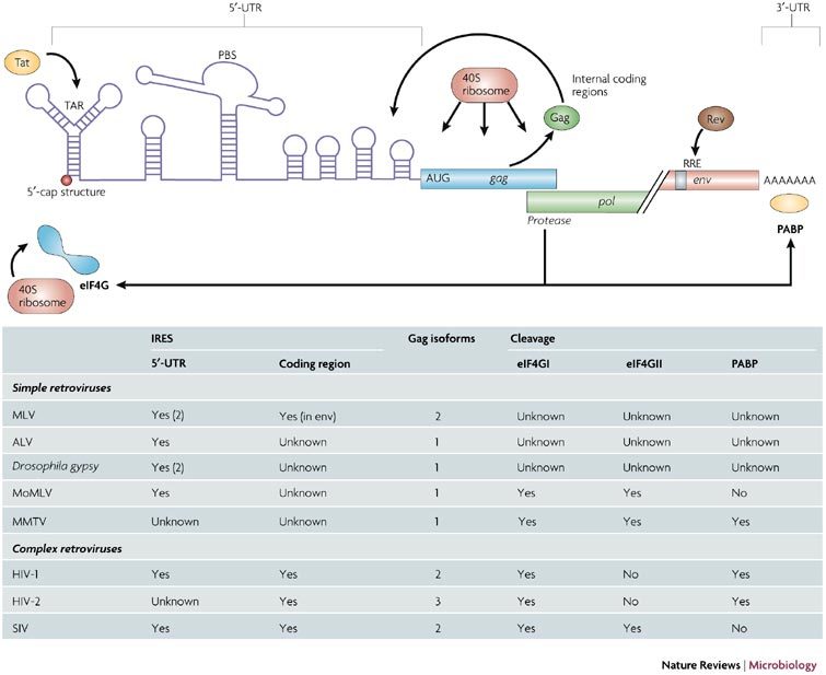
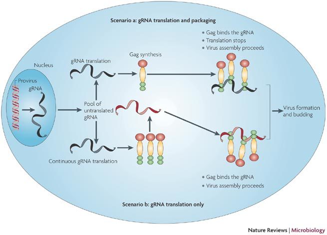
All replication-competent retroviruses contain three main reading frames, gag, pol and env, which are used for the synthesis of structural proteins, enzymes and envelope proteins respectively. Complex retroviruses, such as lentiviruses, also code for regulatory and accessory proteins that have essential roles in viral replication. The concerted expression of these genes ensures the efficient polypeptide production required for the assembly and release of new infectious progeny virions. Retroviral protein synthesis takes place in the cytoplasm and depends exclusively on the translational machinery of the host infected cell. Therefore, not surprisingly, retroviruses have developed RNA structures and strategies to promote robust and efficient expression of viral proteins in a competitive cellular environment.


Conflict of interest statement

The authors declare no competing financial interests.

Figures

Figure 1
Figure 1. An overview of the retroviral life cycle.
The life cycle of retroviruses can be broadly divided into six essential steps, which are shown schematically in the figure: interaction, adsorption and entry into the cell (a); synthesis of reverse transcribed DNA (b); integration (c); transcription, splicing and nuclear export (d); translation and encapsidation (e); and viral assembly and budding (f).
Figure 2
Figure 2. Genetic organization of the 5′-untranslated region (UTR) of the retroviral genomic RNA: the example of SIVmac.
The 5′-UTR (or leader) of the simian immunodeficiency virus (SIV) genomic RNA contains numerous cis-acting sequences, such as the TAR stem-loop, the poly (A) loop, the primer-binding site (PBS), the dimerization initiation site (DIS), the core packaging signals (Psi) and the major splice donor (SD). The R-U5, packaging and coding regions are indicated in the figure.
Figure 3
Figure 3. IRESs in Lentiviruses.
Schematic diagram of the position of the different IRESs within the lentiviral genomic RNAs. The N-truncated Gag isoforms that result from alternative translation initiation from the coding region are shown.
Figure 4
Figure 4. Schematic representation of the secondary structure adopted by the first 450 nucleotides of the HIV-2 Gag coding region.
Secondary structure model deduced from chemical and enzymatic probing data (adapted from Ref. 44). Helices are numbered from P1 to P8, structures after P8 were not named and are not conserved in HIV-1 and simian immunodeficiency virus (SIV)mac. The P2 helix is also absent from HIV-1. The three initiation codons are highlighted in red.
Figure 5
Figure 5. Translational control of lentiviral genomic RNAs.
The figure summarizes the events that modulate and control the translation of the lentiviral genomic RNA. The table summarizes the location of the IRESs, the number of Gag isoforms and the effect of the protease on the translational apparatus for all the retroviruses described in the manuscript. ALV, avian leukosis virus; eIF4G, eukaryotic initiation factor (eIF) 4G (isoform I and II); MLV, murine leukaemia virus; MoMLV, Moloney MLV; MMTV, mouse mammary tumour virus; PABP, poly (A) binding protein; PBS, primer binding site; Rev, regulator of expression of virion proteins; RRE, Rev response element; SIV, simian immunodeficiency virus; TAR, transactivation response element.
Figure 6
Figure 6. Translation and encapsidation of the retroviral genomic RNA.
Transcription of the integrated provirus generates the genome length RNA (gRNA). A fraction of the gRNA is spliced to give the spliced viral RNAs (not shown here). The gRNA is exported to the cytoplasm where it is recruited by active ribosomes (scenarios a and b with gRNA in black) or eventually accumulates as a pool of non-translated RNA (gRNA in red). According to scenario a, the gRNA is translated to give Gag molecules and Gag-Pol (not shown here). Once Gag molecules accumulate, there is a switch from translation to packaging as Gag binds to the gRNA at the 5′-IRES-Psi signal. Virus formation and release by budding follow. In scenario b, the gRNA is continuously translated. Newly formed Gag molecules bind the gRNA from the non-translated pool (red gRNA). Virus formation follows.

Similar articles

Cited by

References

    1. Telenitsky A, Goff SP. Retroviruses. 1997. pp. 121–160.
    1. Paillart JC, Shehu-Xhilaga M, Marquet R, Mak J. Dimerization of retroviral genomes: an inseperable pair. Nature Rev. Mircobiol. 2004;2:461–472. doi: 10.1038/nrmicro903. - DOI - PubMed
    1. Vogt PK. Retroviruses. 1997. pp. 1–25.
    1. Rabson AB, Graves BJ. Retroviruses. 1997. pp. 205–261.
    1. Swanstrom R, Willis JW. Retroviruses. 1997. pp. 263–334.

Publication types