

Review
doi: 10.1007/s00424-006-0194-y.
Epub 2007 Jan 17.
Phototransduction in mouse rods and cones
Affiliations
- PMID: 17226052
- PMCID: PMC2877390
- DOI: 10.1007/s00424-006-0194-y
Item in Clipboard
Review
Phototransduction in mouse rods and cones
Pflugers Arch.
2007 Aug.
Abstract
Phototransduction is the process by which light triggers an electrical signal in a photoreceptor cell. Image-forming vision in vertebrates is mediated by two types of photoreceptors: the rods and the cones. In this review, we provide a summary of the success in which the mouse has served as a vertebrate model for studying rod phototransduction, with respect to both the activation and termination steps. Cones are still not as well-understood as rods partly because it is difficult to work with mouse cones due to their scarcity and fragility. The situation may change, however.
Figures
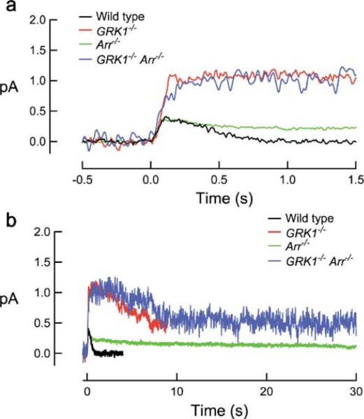
Form of the single-photon response from knockout mouse rods with deficient R* termination on fast (a) and slow (b) timescales. Flashes were delivered at t=0. Figure was kindly supplied by Dr. M. E. Burns
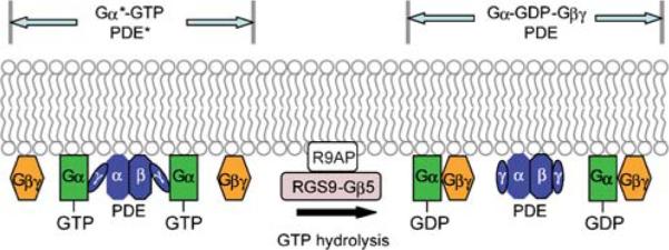
Schematic on the termination G*-PDE*. Gα*-GTP binds to PDEγ and activates PDE. The deactivation of Gα*-GTP is accelerated by the GAP complex, R9AP-RGS9-1-Gβ5L, in which RGS9-1 facilitates the hydrolysis of the bound GTP to GDP, leading to the reformation of the inactive heterotrimeric Gα-GDP-Gβγ and inactive PDE. An asterisk denotes the active state. This step was found to be the rate-limiting step of rod recovery [56]
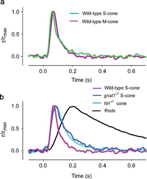
Flash responses of mouse cone photoreceptors from different genotypes. a Comparison of the average response of S-cones to 361-nm flashes and M-cones to 510-nm flashes. b Comparison of the average flash responses to 361-nm flashes of wild-type S-cones, gnat1−/− S-cones, Nrl−/− cones, and rods recorded under the same “OS out” conditions. Each trace is scaled to unity at its peak. Data from Fig. 4e,f of [67]. Reproduced from The Journal of General Physiology, 2006, 127: 359–374. Copyright 2006 The Rockefeller University Press
Similar articles
-
[Physiology of the visual retinal signal: From phototransduction to the visual cycle].J Fr Ophtalmol. 2017 Mar;40(3):239-250. doi: 10.1016/j.jfo.2016.12.006. Epub 2017 Mar 17. J Fr Ophtalmol. 2017. PMID: 28318721 Review. French.
-
Evolutionary transformation of rod photoreceptors in the all-cone retina of a diurnal garter snake.Proc Natl Acad Sci U S A. 2016 Jan 12;113(2):356-61. doi: 10.1073/pnas.1513284113. Epub 2015 Dec 29. Proc Natl Acad Sci U S A. 2016. PMID: 26715746 Free PMC article.
-
Rod and cone photoreceptors: molecular basis of the difference in their physiology.Comp Biochem Physiol A Mol Integr Physiol. 2008 Aug;150(4):369-77. doi: 10.1016/j.cbpa.2008.04.600. Epub 2008 Apr 26. Comp Biochem Physiol A Mol Integr Physiol. 2008. PMID: 18514002 Review.
-
Physiological properties of rod photoreceptor cells in green-sensitive cone pigment knock-in mice.J Gen Physiol. 2007 Jul;130(1):21-40. doi: 10.1085/jgp.200609729. J Gen Physiol. 2007. PMID: 17591985 Free PMC article.
-
Molecular mechanisms characterizing cone photoresponses.Photochem Photobiol. 2007 Jan-Feb;83(1):19-26. doi: 10.1562/2006-02-28-IR-823. Photochem Photobiol. 2007. PMID: 16706600 Review.
Cited by
-
Defective retinal depolarizing bipolar cells in regulators of G protein signaling (RGS) 7 and 11 double null mice.J Biol Chem. 2012 Apr 27;287(18):14873-9. doi: 10.1074/jbc.M112.345751. Epub 2012 Feb 27. J Biol Chem. 2012. PMID: 22371490 Free PMC article.
-
Ophthalmic findings in sheep treated with closantel in Curitiba, Brazil.Vet World. 2020 May;13(5):860-864. doi: 10.14202/vetworld.2020.860-864. Epub 2020 May 8. Vet World. 2020. PMID: 32636579 Free PMC article.
-
Expression Analysis of Visual Arrestin gene during Ocular Development of Olive Flounder (Paralichthys olivaceus).Dev Reprod. 2013 Sep;17(3):231-40. doi: 10.12717/DR.2013.17.3.231. Dev Reprod. 2013. PMID: 25949138 Free PMC article.
-
A visually guided swim assay for mouse models of human retinal disease recapitulates the multi-luminance mobility test in humans.Saudi J Ophthalmol. 2023 Nov 18;37(4):313-320. doi: 10.4103/sjopt.sjopt_155_23. eCollection 2023 Oct-Dec. Saudi J Ophthalmol. 2023. PMID: 38155679 Free PMC article.
-
Origin and control of the dominant time constant of salamander cone photoreceptors.J Gen Physiol. 2012 Aug;140(2):219-33. doi: 10.1085/jgp.201110762. Epub 2012 Jul 16. J Gen Physiol. 2012. PMID: 22802362 Free PMC article.
References
-
- Carter-Dawson LD, LaVail MM. Rods and cones in the mouse retina. I. Structural analysis using light and electron microscopy. J Comp Neurol. 1979;188:245–262. - PubMed
-
- Humphries MM, Rancourt D, Farrar GJ, Kenna P, Hazel M, Bush RA, Sieving PA, Sheils DM, McNally N, Creighton P, et al. Retinopathy induced in mice by targeted disruption of the rhodopsin gene. Nat Genet. 1997;15:216–219. - PubMed
Publication types
MeSH terms
Substances
Grants and funding
LinkOut - more resources
Full Text Sources
Other Literature Sources
