Review
. 2007 Feb;29(2):120-32.
doi: 10.1002/bies.20526.

Planar cell polarity signaling in vertebrates

Affiliations
Review

Planar cell polarity signaling in vertebrates

Chonnettia Jones et al. Bioessays. 2007 Feb.

Abstract

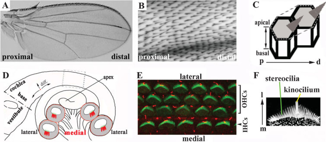
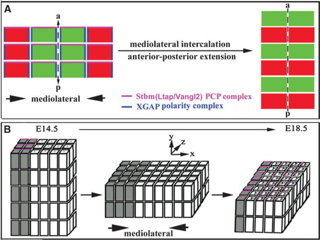
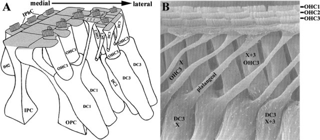
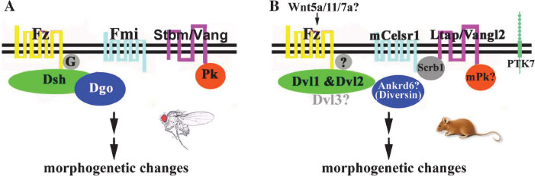
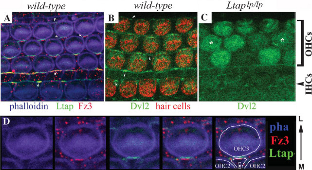
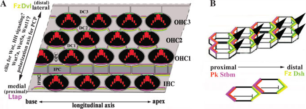
Planar cell polarity (PCP) refers to the polarization of a field of cells within the plane of a cell sheet. This form of polarization is required for diverse cellular processes in vertebrates, including convergent extension (CE), the establishment of PCP in epithelial tissues and ciliogenesis. Perhaps the most distinct example of vertebrate PCP is the uniform orientation of stereociliary bundles at the apices of sensory hair cells in the mammalian auditory sensory organ. The establishment of PCP in the mammalian cochlea occurs concurrently with CE in this ciliated epithelium, therefore linking three cellular processes regulated by the vertebrate PCP pathway in the same tissue and emerging as a model system for dissecting PCP signaling. This review summarizes the morphogenesis of this model system to assist the interpretation of the emerging data and proposes molecular mechanisms underlying PCP signaling in vertebrates.


Figures

Figure 1
Figure 1. Planar cell polarity
A–C: PCP in the Drosophila wing. The bristles point from the proximal to distal direction and are uniformly oriented (A,B). The PCP displayed by the Drosophila wing is illustrated diagrammatically (C). Note that the uniform orientation of bristles manifests a polarity along the proximal-to-distal (p–d) axis of the plane formed by the epithelial sheet that is perpendicular to the basal–apical axis of the epithelium. D: A diagram of the cochlea. The cochlear duct forms a spiral and the tip of the cochlea, or apex, lies distally from the base of the cochlea, which is juxtaposed to the vestibule. (Note that the terms ‘‘apex’’ and ‘‘base’’ used for the cochlea are different from the terms used to describe the apical and basolateral domains of individual epithelial cells.) The longitudinal axis is along the length of the cochlear duct, while the mediolateral axis is from the center to the periphery of the cochlea. Shown in grey are cross-sections of the cochlear duct, illustrating the positions of the sensory hair cells (red) and the stereocilia bundles (green) protruding from their apical surfaces into the lumen of the duct. The gray dotted box indicates the region highlighted in panel E. E: The organ of Corti viewed from its surface in a confocal image. The stereociliary bundles (green) on the apical surface of each hair cell are patterned into a ‘‘V’’-shaped structure and all the ‘‘V’’s are uniformly oriented, showing a distinctive polarity that is parallel to the sensory epithelial sheet along the mediolateral axis. The kinocilia (red) are positioned at the vertices of the ‘‘V’’-shaped stereocilia. Note that the entire organ of Corti is ciliated. Some of the cilia (red) are from supporting cells (not shown). The single row of inner hair cells (IHCs) is located toward the center (medial) of the cochlear duct ,while the outer hair cells (OHCs) are located lateral to the inner hair cells toward the periphery of the cochlear duct. F: Stereocilia and a kinocilium of one hair cell, showing the stereociliary bundles arranged in a ‘‘V’’ shape with a kinocilium-positioned at the vertex of the V. m: medial; l: lateral.
Figure 2
Figure 2. Convergent extension
A: A schematic illustration of CE during gastrulation and neurulation in Xenopus and zebrafish. Cells project mediolaterally oriented lamellipodia and exert traction along the mediolateral axis to drive convergence along the same axis and extension along the perpendicular axis (anterior–posterior). The PCP complex of Stbm and PK (magenta) are thought to be at the anterior ends of the cells during CE, and the XGAP polarity complex (blue) are localized to the mediolateral ends of the cells, orienting the cells at both axes for polarized cellular rearrangement. a, anterior; p, posterior. B: The proposed CE in the cochlea during terminal differentiation of the organ of Corti. The mature organ of Corti is derived from the shorter and thicker ‘‘primordial domain’’ (white boxes). Cells of the primordial domain are evidenced by a zone of non-proliferating cells around E13.5–E14.5 in mice. In PCP mutants, the organ of Corti is widened (X-axis) and shortened (Z-axis), suggesting that the developing organ of Corti undergoes mediolateral intercalation. While the organ of Corti is postmitotic after E13.5–E14.5, the cells adjacent to the developing organ of Corti (shaded boxes) continue to divide and show a distinct polarized localization of Ltap/Vangl2/Stbm (pink) at E14.5 and E18.5 along the extension axis. In the developing organ of Corti (white boxes), Ltap/Vangl2 (pink) apparently is localized to the medial end of hair cells, perpendicular to the polarized localization of Ltap/Vangl2 in the adjacent region (shaded boxes). The significance of the polarized subcellular localization of PCP proteins in the region medial to the organ of Corti has not been investigated. During the extension of the cochlea, the kinocilia (green) are displaced toward the lateral sides of the hair cells and the stereocilia develop and acquire the ‘‘V’’-shaped structure. The morphology of the developing cochlea and the localization of PCP proteins during the extension from E14.5–E18.5 are dynamic and have not been carefully characterized.
Figure 3
Figure 3. The polarity of supporting cells of the organ of Corti
A: A diagram of the cellular architecture of the organ of Corti showing the intricate cell-cell contacts between hair cells and supporting cells and among the different types of supporting cells. The polarity of the phalangeal processes of supporting cells is also illustrated. The IHCs are separated from each other by the IPhCs at the apical surface. The IHCs are separated from the OHCs by the IPCs and OPCs. The OHCs within each row are separated by the phalangeal processes of the OPCs and the first and second rows of Deiters cells (DC1 and DC2). The phalangeal processes of the third row of DCs (DC3) form the lateral border for the sensory hair cells at the apical surface. IPhC, inner phalangeal cell; IPC, inner pillar cell; OPC, outer pillar cell; DC, Deiters cell; IHC, inner hair cell; OHC, outer hair cell. B: A scanning electron microscope image viewed from the lateral side of the organ of Corti to illustrate the polarity of the phalangeal processes of supporting cells around the third row of OHCs (OHC3). The hair cells (marked with OHC1, OHC2 and OHC3) at the surface are also visible. The phalangeal processes of the supporting cells project in precise orientations toward the lumen of the cochlear epithelium to separate hair cells from each other. For instance, a Deiters cell (DC3) at position X along the longitudinal axis projects its phalangeal processes toward the lateral direction to border the third row of outer hair cells at position X + 3 (toward the apex of the cochlear duct). (Panel A is modified from an illustration by Slepecky, 1996, with permission from Springer. Scanning electron micrograph in B is provided by Dr. Y. Raphael, University of Michigan, Ann Arbor, MI. From Raphael et al. 1991, reprinted by permission of John Wiley and Sons, Inc.).
Figure 4
Figure 4. The PCP pathway
A: Drosophila PCP pathway, showing the core PCP complexes of Fz–Dsh–Dgo and Stbm–Pk. Fmi is likely to be associated with both complexes. B: Vertebrate PCP pathway, showing additional components (PTK7 and Scribble) in PCP regulation in mice. The proposed interactions among the vertebrate PCP components are based on available data (see text). The association of Scrb1 with mCelsr1 is based on the ubiquitous localization of Scrb1 to apical cell membranes in the organ of Corti. The role of mouse Ankrd6, Pk and Dvl3 in PCP has not been demonstrated, and the role of Wnts in establishing epithelial PCP has yet to be tested.
Figure 5
Figure 5. Polarized localization of Ltap/Vangl2, Dvl2 and Fz3 in the organ of Corti
A: A whole-mount E17.5 organ of Corti that expresses Ltap–GFP(green) (D. Qian and P. Chen, unpublished data)was stained with an antibody against Fz3 (red). The tissue was also stained with phalloidin (blue) to outline the hair cells. Both Ltap–GFP and Fz3 appear to be on the medial side of the hair cells. However, non-overlapping localization of Ltap–GFP and Fz3 is observed. The Ltap–GFP signal is slightly lateral to that of Fz3. The arrowheads indicate the localization of Ltap–GFP and Fz3 to boundaries formed between supporting cells. B: Dvl2–GFP localization in the organ of Corti, showing an apparent localization (green) to the lateral end of the hair cells (red) at the apical surface of the organ of Corti. C: The polarized localization of Dvl2 is altered in PCP mutant LtapLp/Lp mice. The asterisks indicate two hair cells with altered membrane association of Dvl2–GFP. In the other hair cells, no enrichment of Dvl2 in the cell membrane was observed. The apical cytoplasmic GFP signal seems to intensify in some of the hair cells. Interestingly, the membrane association of Dvl2–GFP in the supporting cells that separate inner from outer hair cells appears to remain, albeit less intense. D: A large view of Fz3 (red) and Ltap–GFP (green) localization at the boundaries formed between a hair cell (OHC3) and a supporting cell (DC1) as in (A). Phalloidin (blue) outlines the cortex of cells and the V-shaped stereocilia on OHC3. Note that Fz3 is slightly lateral to Ltap–GFP at the boundary and may be contributed by DC1. M: medial; L: lateral.
Figure 6
Figure 6. PCP signaling in vertebrates
A: A schematic drawing of the PCP regulation in the organ of Corti. The cellular interfaces of the organ of Corti are outlined. In this model, we tentatively placed Fz3 (yellow) on the lateral side of supporting cells (grey) and proposed that Fz3 recruits Dvl2 (green) to the same location. In the hair cells (black), Dvl2 is also localized to the lateral side. The mechanism for recruiting Dvl2 to the lateral side of the hair cells is not known. Ltap/Vangl2 (pink) is localized to the medial side of hair cells and supporting cells. The interaction between the extracellular domains of Ltap and Fz3 from the two adjacent cells enforces the polarization of PCP complexes in the organ of Corti. The detailed assignment of the PCP proteins to the individual side of the cells is based on experimental data in vertebrates and demonstrated mechanisms for fly PCP (B) regulation, but not definitive. Vertebrates may use completely different mechanisms for polarization of PCP complexes. The identity and the role of mouse Pk in PCP regulation in the organ of Corti is not known. A critical test of the proposed model is to determine whether mPk proteins play a similar antagonistic role for Dvl localization in the cochlea. The upstream factors involve Wnts, possibly including Wnt7a, Wnt5a, and Wnt11.The exact role of cilia in PCP regulation is not clear. They may be involved in direct signal transduction or in downstream effector gene functions. Since cilia are implicated in PCP signaling, it is possible that the signaling pathways mediated by cilia, such as Hedgehog pathway, may also be involved in PCP signaling. B: PCP regulation in Drosophila. Pk (orange) is recruited to the membrane by Stbm (pink) and inhibits the localization of Dsh (green) to the same site. The antagonist role of Pk to Dsh restricts Dsh/Fz complex to the opposite side of the cells. The interactions between the extracellular domains of Stbm and Fz (yellow) at the interface of the two adjacent cells stabilize the polarized localization of Stbm/Pk and Fz/Dsh to the opposite sides of the cells.

References

    1. Gubb D, Garcia-Bellido A. A genetic analysis of the determination of cuticular polarity during development in Drosophila melanogaster. J Embryol Exp Morphol. 1982;68:37–57. - PubMed
    1. Strutt H, Strutt D. Long-range coordination of planar polarity in Drosophila. Bioessays. 2005;27:1218–1227. - PubMed
    1. Klein TJ, Mlodzik M. Planar cell polarization: an emerging model points in the right direction. Annu Rev Cell Dev Biol. 2005;21:155–176. - PubMed
    1. Wallingford JB, Rowning BA, Vogeli KM, Rothbacher U, Fraser SE, Harland RM. Dishevelled controls cell polarity during Xenopus gastrulation. Nature. 2000;405:81–85. - PubMed
    1. Carreira-Barbosa F, Concha ML, Takeuchi M, Ueno N, Wilson SW, et al. Prickle 1 regulates cell movements during gastrulation and neuronal migration in zebrafish. Development. 2003;130:4037–4046. - PubMed