

Impact of endoscopic ultrasound-guided fine needle biopsy for diagnosis of pancreatic masses
- PMID: 17226911
- PMCID: PMC4065960
- DOI: 10.3748/wjg.v13.i2.289
Impact of endoscopic ultrasound-guided fine needle biopsy for diagnosis of pancreatic masses
Abstract
Aim: To evaluate the diagnostic accuracy of histological evaluation of pancreatic tissue samples obtained by a modified method for recovering and processing the endoscopic ultrasound (EUS)-guided fine needle aspiration (FNA) material in the differential diagnosis of pancreatic solid masses.
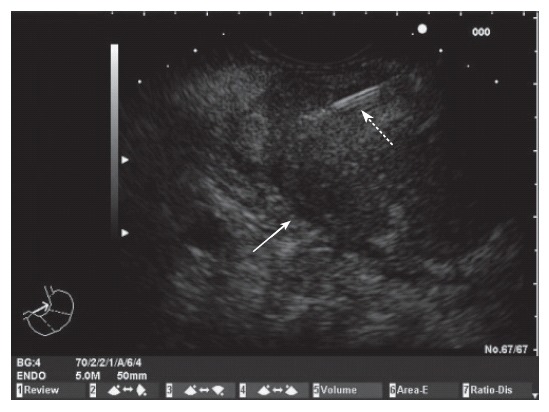
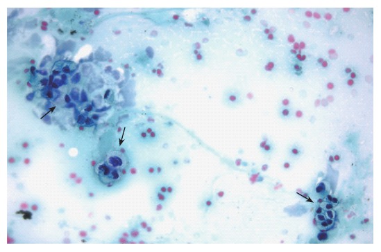
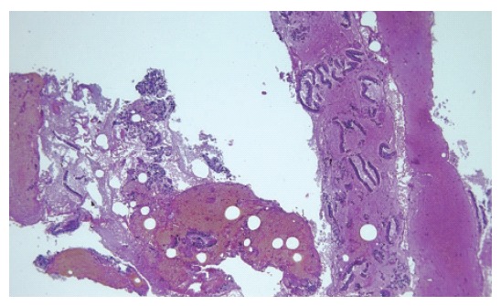
Methods: Sixty-two consecutive patients with pancreatic masses were prospectively studied. EUS was performed by the linear scanning Pentax FG-38UX echoendoscope. Three FNAs (22G needle) were carried out during each procedure. The materials obtained with first and second punctures were processed for cytological study. Materials of the third puncture were recovered into 10% formol solution by careful injection of saline solution through the needle, and processed for histological study.
Results: Length of the core specimen obtained for histological analysis was 6.5 +/- 5.3 mm (range 1-22 mm). Cytological and histological samples were considered as adequate in 51 (82.3%) and 52 cases (83.9%), respectively. Overall sensitivity of both pancreatic cytology and histology for diagnosis of malignancy was 68.4%. Contrary to cytology, histology was able to diagnose tumours other than adenocarcinomas, and all cases of inflammatory masses. Combination of cytology and histology allowed obtaining an adequate sample in 56 cases (90.3%), with a global sensitivity of 84.21%, specificity of 100% and an overall accuracy of 90.32%. The complication rate was 1.6%.
Conclusion: Adequate pancreatic core specimens for histological examination can be obtained by EUS-guided FNA. This technique is mainly useful for the diagnosis of different types of pancreatic tumours and evaluation of benign diseases.
Figures

References
-
- Tamm E, Charnsangavej C. Pancreatic cancer: current concepts in imaging for diagnosis and staging. Cancer J. 2001;7:298–311. - PubMed
-
- Cohen SJ, Pinover WH, Watson JC, Meropol NJ. Pancreatic cancer. Curr Treat Options Oncol. 2000;1:375–386. - PubMed
-
- Rösch T. Endoscopic ultrasonography. Br J Surg. 1997;84:1329–1331. - PubMed
-
- Hawes RH. Endoscopic ultrasound. Gastrointest Endosc Clin N Am. 2000;10:161–174, viii. - PubMed
-
- Ribeiro A, Vazquez-Sequeiros E, Wiersema LM, Wang KK, Clain JE, Wiersema MJ. EUS-guided fine-needle aspiration combined with flow cytometry and immunocytochemistry in the diagnosis of lymphoma. Gastrointest Endosc. 2001;53:485–491. - PubMed
Publication types
MeSH terms
LinkOut - more resources
Full Text Sources
Medical
