

Gallbladder lymphangioma: a case report and review of the literature
- PMID: 17226918
- PMCID: PMC4065967
- DOI: 10.3748/wjg.v13.i2.320
Gallbladder lymphangioma: a case report and review of the literature
Abstract
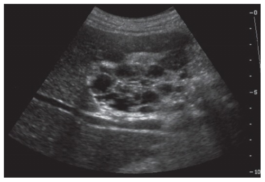
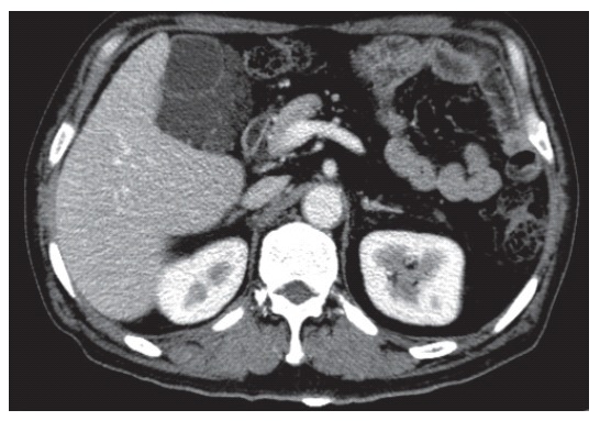
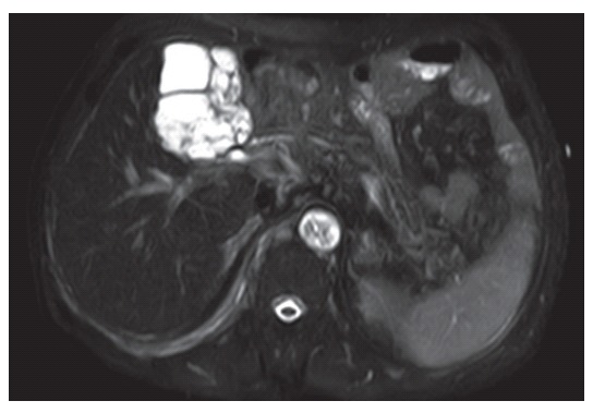
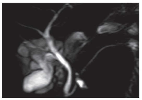
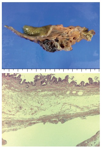
Lymphangiomas are rare, benign tumors of the lymphatic system, usually present in children aged 5 years and younger. Because they are asymptomatic until the mass enlarges to cause symptoms, most lymphangiomas are diagnosed at adulthood incidentally. We experienced a case of a 60-year-old man diagnosed with a cystic lymphangioma of the gallbladder, which was successfully resected without any complication. Magnetic resonance imaging and magnetic resonance cholangiopancreatography were very helpful for the diagnosis of the cystic lesion around the gallbladder as were ultrasonography and computed tomography scan. These showed a multi-lobulated cystic mass with intact cystic duct and bile duct in the gallbladder fossa. The patient underwent an open cholecystectomy and the histological findings were consistent with a cystic lymphangioma of the gallbladder. We here report the case of cystic lymphangioma of the gallbladder with a review of the literature.
Figures
Similar articles
-
Gallbladder lymphangioma: a case report and review of the literature.J Hepatobiliary Pancreat Surg. 2005;12(5):405-8. doi: 10.1007/s00534-005-0997-9. J Hepatobiliary Pancreat Surg. 2005. PMID: 16258810
-
Cystic lymphangioma of the gallbladder: report of a case.Surg Today. 2008;38(1):81-4. doi: 10.1007/s00595-007-3564-y. Epub 2007 Dec 24. Surg Today. 2008. PMID: 18085372
-
Gallbladder lymphangioma: MR findings.Abdom Imaging. 2002 Jan-Feb;27(1):54-7. doi: 10.1007/s00261-001-0051-6. Abdom Imaging. 2002. PMID: 11740609
-
Gall-bladder and hepatoduodenal ligament lymphangioma - case report and literature review.Pol Przegl Chir. 2013 Jan;85(1):39-43. doi: 10.2478/pjs-2013-0007. Pol Przegl Chir. 2013. PMID: 23509201 Review.
-
Lymphangioma of the kidney.Int J Urol. 2002 Mar;9(3):178-82. doi: 10.1046/j.1442-2042.2002.00437.x. Int J Urol. 2002. PMID: 12010331 Review.
Cited by
-
Extensive and invasive lymphangioma circumscriptum in a young female: A rare case report and review of the literature.Indian Dermatol Online J. 2013 Jul;4(3):199-201. doi: 10.4103/2229-5178.115516. Indian Dermatol Online J. 2013. PMID: 23984233 Free PMC article.
-
Intracystic papillary neoplasm with an associated mucinous adenocarcinoma arising in Rokitansky-Aschoff sinus of the gallbladder.Surg Case Rep. 2016 Dec;2(1):62. doi: 10.1186/s40792-016-0189-7. Epub 2016 Jun 18. Surg Case Rep. 2016. PMID: 27316722 Free PMC article.
-
Lymphangioma of the gallbladder in adults: review of the literature and a case report.J Gastrointest Surg. 2012 Mar;16(3):663-8. doi: 10.1007/s11605-011-1770-9. Epub 2011 Nov 4. J Gastrointest Surg. 2012. PMID: 22052109
-
A comprehensive exploration of gallbladder health: from common to rare imaging findings.Abdom Radiol (NY). 2025 Jan;50(1):131-151. doi: 10.1007/s00261-024-04431-4. Epub 2024 Jul 2. Abdom Radiol (NY). 2025. PMID: 38953999 Review.
-
Extensive congenital vulvar lymphangioma mimicking genital warts.Indian J Dermatol. 2010;55(1):121-2. doi: 10.4103/0019-5154.60372. Indian J Dermatol. 2010. PMID: 20418997 Free PMC article. No abstract available.
References
-
- Noh KW, Bouras EP, Bridges MD, Nakhleh RE, Nguyen JH. Gallbladder lymphangioma: a case report and review of the literature. J Hepatobiliary Pancreat Surg. 2005;12:405–408. - PubMed
-
- Ohba K, Sugauchi F, Orito E, Suzuki K, Ohno T, Mizoguchi N, Koide T, Terashima H, Nakano T, Mizokami M. Cystic lymphangioma of the gall-bladder: a case report. J Gastroenterol Hepatol. 1995;10:693–696. - PubMed
-
- Lörken M, Marnitz U, Manegold E, Schumpelick V. Intra-abdominal lymphangioma. Chirurg. 2001;72:72–77. - PubMed
-
- Bishop MD, Steer M. Pancreatic cystic lymphangioma in an adult. Pancreas. 2001;22:101–102. - PubMed
-
- Takiff H, Calabria R, Yin L, Stabile BE. Mesenteric cysts and intra-abdominal cystic lymphangiomas. Arch Surg. 1985;120:1266–1269. - PubMed
Publication types
MeSH terms
LinkOut - more resources
Full Text Sources
Medical
