. 2007 Jan 16:6:6.
doi: 10.1186/1476-4598-6-6.

Breast cancer metastasis suppressor 1 (BRMS1) inhibits osteopontin transcription by abrogating NF-kappaB activation

Affiliations

Breast cancer metastasis suppressor 1 (BRMS1) inhibits osteopontin transcription by abrogating NF-kappaB activation

Rajeev S Samant et al. Mol Cancer. .

Abstract

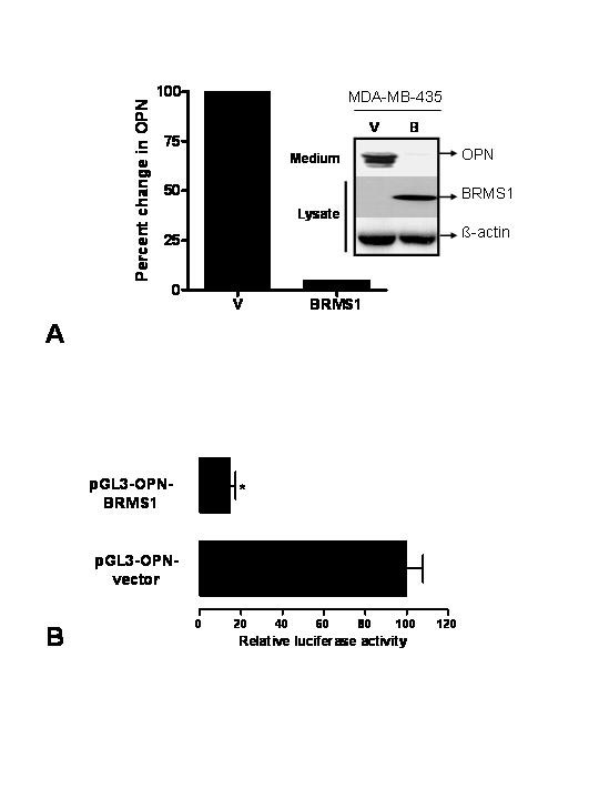
Background: Osteopontin (OPN), a secreted phosphoglycoprotein, has been strongly associated with tumor progression and aggressive cancers. MDA-MB-435 cells secrete very high levels of OPN. However metastasis-suppressed MDA-MB-435 cells, which were transfected with breast cancer metastasis suppressor 1 (BRMS1), expressed significantly less OPN. BRMS1 is a member of mSin3-HDAC transcription co-repressor complex and has been shown to suppress the metastasis of breast cancer and melanoma cells in animal models. Hence we hypothesized that BRMS1 regulates OPN expression.

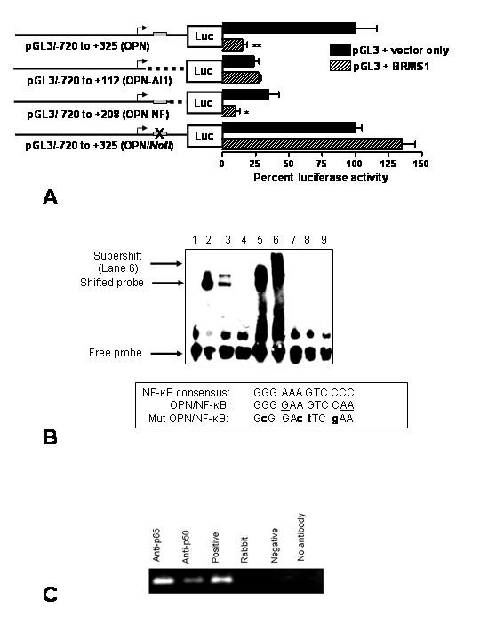
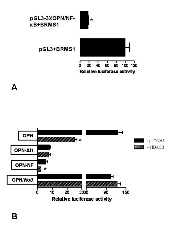
Results: The search for a BRMS1-regulated site on the OPN promoter, using luciferase reporter assays of the promoter deletions, identified a novel NF-kappaB site (OPN/NF-kappaB). Electrophoretic mobility shift assays and chromatin immunoprecipitations (ChIP) confirmed this site to be an NF-kappaB-binding site. We also show a role of HDAC3 in suppression of OPN via OPN/NF-kappaB.

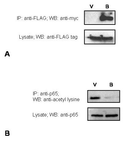
Conclusion: Our results show that BRMS1 regulates OPN transcription by abrogating NF-kappaB activation. Thus, we identify OPN, a tumor-metastasis activator, as a crucial downstream target of BRMS1. Suppression of OPN may be one of the possible underlying mechanisms of BRMS1-dependent suppression of tumor metastasis.


Figures

Figure 1
Figure 1
A. Expression of OPN is down-regulated by 95% in the 435/BRMS1 cells. Serum-free conditioned medium from equal numbers of pcDNA transfected MDA-MB-435 (V) and 901-BRMS1-transfected 435 cells, 435/BRMS1 (BRMS1) was resolved using a 12.5% SDS-PAGE, transferred to a PVDF membrane and probed with the anti-human OPN monoclonal antibody [30]. The bar graph depicts the percent change in OPN expression based on densitometric analysis of the immunoblot. To confirm the BRMS1 expression in these cells, the cells were lysed in NP-40 lysis buffer and 30 ug of protein was resolved using a 12.5% SDS-PAGE, transferred to a PVDF membrane and probed with the anti-901 monoclonal antibody (for BRMS1 epitope). The membrane was reprobed for levels of β-actin to confirm equal loading. B. BRMS1 suppresses activity of the human OPN promoter. COS-7 cells were co-transfected with pGL3-OPN [28] and pCMV-myc or pCMV-myc-BRMS1 using Lipofectamine 2000 (Invitrogen). Luciferase activity was normalized to the total protein concentration. Data is expressed as Relative luciferase activity, where control is 100%. The data represents five independent experiments in triplicate. * indicates significant suppression (p < 0.05).
Figure 2
Figure 2
A. The OPN promoter lacking the NF-κB site is relieved for BRMS1 suppression. The grey box represents the predicted NF-κB site. The dotted line indicates the region deleted in the construct. OPN-ΔI1 does not have the NF-κB-binding site and is not suppressed by BRMS1. OPN/NotI, in which the NF-κB site is abolished by inserting a NotI site in its place, is refractory to suppression by BRMS1. Data is expressed as Relative luciferase activity of control, where control (pGL3-OPN) is 100%. The data shown represents more than three independent experiments in triplicate. * indicates significant suppression, p value < 0.05 and ** indicates p < 0.01 compared to respective controls. B. Recombinant p65 and p50 bind to and retard the mobility of the predicted NF-κB site from the promoter of OPN. Lane 1: Probe bearing the NF-κB site from OPN promoter; Lane 2: Probe + recombinant p50; Lane 3: Probe + nuclear extract of MDA-MB-435; Lane 4: Probe + unlabeled 'cold' consensus probe + Nuclear extract of MDA-MB-435; Lane 5: Probe + recombinant p65; Lane 6: Probe + recombinant p65 + anti-p65 antibody; Lane 7: Mutant probe; Lane 8: Mutant Probe + recombinant p65; Lane 9: Mutant Probe + recombinant p50. The box gives the sequence comparison of the OPN/NF-κB with the consensus NF-κB-binding site. The underscored bases in OPN/NF-κB represent variation from the consensus. Mutant OPN/NF-κB (Mut OPN/NF-κB) is also shown with the mutations represented in bold lower case. C. NF-κB subunits bind to the OPN promoter in vivo. MDA-MB-435 cells were fixed with formaldehyde, lysed, and then sonicated. In vivo cross-linked chromatin was then precipitated independently using p65 (Anti-p65), p50 (Anti-p50), normal rabbit IgG (Rabbit), no antibody, or Positive and Negative kit control antibodies (Active Motif). The recovered immunoprecipitated DNA was then used for PCR with primers specific for the OPN/NF-κB-containing promoter segment. A 151 bp product corresponding to a region 1575 bp upstream of the OPN/NF-kB site (that lacks a predicted NF-κB site [12]) was absent in a control PCR following ChIP Primers: 5'-TTCCCCCTACCAAATGTTCA-3' and 5'-TGCTGCAAAAGTAATTGTGGTT-3'.
Figure 3
Figure 3
A. BRMS1 downregulates OPN via the NF-κB site. A reporter plasmid bearing three NF-κB sites from the OPN promoter, pGL3-3XOPN/NF-κB, upstream of the SV40 promoter or pGL3-control was co-transfected with pCMV-mycBRMS1 into COS-7 cells. Luciferase activity from this reporter construct was measured. The experiment was repeated thrice in triplicate.* indicates significant suppression compared with respective controls (p < 0.05). B. The OPN promoter lacking an intact NF-κB site is relieved for suppression by HDAC3. The OPN promoter construct, pGL3-OPN, and its deletions, OPN-NF (retains the NF-κB-binding site) and OPN-ΔI1 (lacks the NF-κB-binding site) and the OPN/NotI construct (OPN/NF-κB site is replaced with a NotI site) were co-transfected with pcDNA3 or pcDNA3-FLAG-HDAC3 in COS-7 cells and monitored for the effect of HDAC3 on the promoter activity of OPN. The results shown represent the experiment done twice in triplicate. * indicates significant suppression compared with respective controls (p < 0.05).
Figure 4
Figure 4
A. BRMS1 co-immunoprecipitates HDAC3. FLAG-HDAC3 was co-transfected into COS-7 cells with either pCMV-myc (V) or pCMV-myc-BRMS1(B) using Lipofectamine 2000 (Invitrogen). The cell lysate was immunoprecipitated with 1 μg anti-FLAG Ab (Sigma). The precipitated proteins were resolved by SDS-PAGE and immunoblotted with 1:500 dilution anti-myc Ab (BD Clontech). Independently the cell lysate was immunoblotted with anti-FLAG antibody to confirm expression of FLAG-HDAC3. B. BRMS1 reduces acetylation of p65. MDA-MB-435 cells were transfected with pCMV-myc (V) or pCMV-myc-BRMS1 (B) and immunoprecipitated with anti-p65 antibody. The immunoprecipitate was resolved on SDS-PAGE, transferred to PVDF membrane and immunoblotted with anti-acetyl lysine antibody. The lysate shows that the levels of p65 are not altered by BRMS1.

Similar articles

Cited by

References

    1. Singhal H, Bautista DS, Tonkin KS, O'Malley FP, Tuck AB, Chambers AF, Harris JF. Elevated plasma osteopontin in metastatic breast cancer associated with increased tumor burden and decreased survival. Clin Cancer Res. 1997;3:605–611. - PubMed
    1. Tuck AB, O'Malley FP, Singhal H, Harris JF, Tonkin KS, Kerkvliet N, Saad Z, Doig GS, Chambers AF. Osteopontin expression in a group of lymph node negative breast cancer patients. Int J Cancer. 1998;79:502–508. doi: 10.1002/(SICI)1097-0215(19981023)79:5<502::AID-IJC10>3.0.CO;2-3. - DOI - PubMed
    1. Rudland PS, Platt-Higgins A, El-Tanani M, De Silva Rudland S, Barraclough R, Winstanley JH, Howitt R, West CR. Prognostic significance of the metastasis-associated protein osteopontin in human breast cancer. Cancer Res. 2002;62:3417–3427. - PubMed
    1. Coppola D, Szabo M, Boulware D, Muraca P, Alsarraj M, Chambers AF, Yeatman TJ. Correlation of osteopontin protein expression and pathological stage across a wide variety of tumor histologies. Clin Cancer Res. 2004;10:184–190. doi: 10.1158/1078-0432.CCR-1405-2. - DOI - PubMed
    1. Cook AC, Tuck AB, McCarthy S, Turner JG, Irby RB, Bloom GC, Yeatman TJ, Chambers AF. Osteopontin induces multiple changes in gene expression that reflect the six "hallmarks of cancer" in a model of breast cancer progression. Mol Carcinog. 2005;43:225–236. doi: 10.1002/mc.20105. - DOI - PubMed

Publication types

MeSH terms