. 2007 Jan 23;104(4):1189-94.
doi: 10.1073/pnas.0610297104. Epub 2007 Jan 16.

Structural analysis and dimerization potential of the human TAF5 subunit of TFIID

Affiliations

Structural analysis and dimerization potential of the human TAF5 subunit of TFIID

Suparna Bhattacharya et al. Proc Natl Acad Sci U S A. .

Abstract

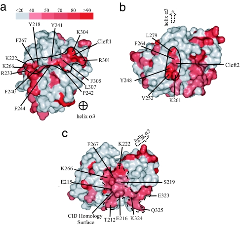
TFIID is an essential factor required for RNA polymerase II transcription but remains poorly understood because of its intrinsic complexity. Human TAF5, a 100-kDa subunit of general transcription factor TFIID, is an essential gene and plays a critical role in assembling the 1.2 MDa TFIID complex. We report here a structural analysis of the TAF5 protein. Our structure at 2.2-A resolution of the TAF5-NTD2 domain reveals an alpha-helical domain with distant structural similarity to RNA polymerase II CTD interacting factors. The TAF5-NTD2 domain contains several conserved clefts likely to be critical for TFIID complex assembly. Our biochemical analysis of the human TAF5 protein demonstrates the ability of the N-terminal half of the TAF5 gene to form a flexible, extended dimer, a key property required for the assembly of the TFIID complex.


Conflict of interest statement

The authors declare no conflict of interest.

Figures

Fig. 1.
Fig. 1.
Primary sequence organization of hTAF5. Schematic diagram of the domain structure present in human TAF5 where NTD1/LisH corresponds to LIS1 homology domain (29), NTD2 corresponds to the α-helical domain reported here, and WD40 repeats are predicted to form a closed β-propeller structure.
Fig. 2.
Fig. 2.
Structure of the hTAF5-NTD2 domain. (a) Diagram of the hTAF5-NTD2. (b) Top view of the α-helical domain of the hTAF5-NTD2 showing the arrangement of the other helices around the central helix (α3) in the crystal structure. All of the helices and the β strands are labeled. Two views in a and b are related by rotation of 90° around a horizontal axis. (c) Topology diagram of the secondary structural elements of the hTAF5-NTD2 domain to show the arrangement of the helical bundle (front view) and the helical sheet (back view) of the crystal structure.
Fig. 3.
Fig. 3.
Conserved surface features of the TAF-NTD2 domain. The surface conservation score was calculated with ESpript (28) based on the sequence alignment in SI Fig. 7 and was mapped on the hTAF5-NTD2 molecular surface by PYMOL. The shade of red in the figure indicates the degree of conservation within TAF5 family members (white, no conservation, to dark red, absolute conservation). The scale bar (a) in the figure indicates the degree of residue conservation from 0.0 to 1.0 conserved cleft 1, the largest involuted cleft on the TAF5-NTD2 domain with conserved residues. Helix α3 is pointed out of the plane of the page. (b) Conserved cleft 2. (c) Conserved residue distributions on the surface of hTAF5-NTD2 corresponding to the CID homology surface.
Fig. 4.
Fig. 4.
Structural similarity between TAF5-NTD2 and CID domains. Shown is a stereo diagram of the core helical bundle (α2–α5) from hTAF5-NTD2 superimposed onto the core helices from the PCF11 structure responsible for CTD interaction. hTAF5-NTD2 is shown in red, whereas PCF11 is shown in blue.
Fig. 5.
Fig. 5.
Biophysical characterization of the association state of TAF5 fragments. Shown is an estimation of native molecular mass by SEC-LS. The molecular mass from the SEC-LS study was estimated to be 55.7 ± 2.8 kDa, confirming the presence of the dimeric form of the hTAF5-NTD1-NTD2 fragment in the solution in buffer containing no divalent cations near physiologic ion strength.
Fig. 6.
Fig. 6.
Hypothetical model for the organization of the TAF5-NTD1 and -NTD2 domains within TFIID. Fig. 6 is adapted from figure 6 of Leurent et al. (21), which roughly mapped the relative TAF positions by immunomapping studies. The TAF5-NTD1 domain has been modeled by using the LisH domain from Lis1 (31), whereas the NTD2 domain has been determined in this study. The 30-Å linker between the C terminus of the LisH region of the NTD1 domain and the N terminus of the NTD2 domain is a rough estimate of the distance between the domains spanned by 70 residues in the human sequence and is presumably flexible.

References

    1. Lee TI, Causton HC, Holstege FC, Shen WC, Hannett N, Jennings EG, Winston F, Green MR, Young RA. Nature. 2000;405:701–704. - PubMed
    1. Tora L. Genes Dev. 2002;16:673–675. - PubMed
    1. Chalkley GE, Verrijzer CP. EMBO J. 1999;18:4835–4845. - PMC - PubMed
    1. Verrijzer CP, Yokomori K, Chen JL, Tjian R. Science. 1994;264:933–941. - PubMed
    1. Verrijzer CP, Chen JL, Yokomori K, Tjian R. Cell. 1995;81:1115–1125. - PubMed

Publication types

Substances

Associated data