. 2007 Jan 23;104(4):1366-70.
doi: 10.1073/pnas.0609848104. Epub 2007 Jan 16.

Histoplasma capsulatum alpha-(1,3)-glucan blocks innate immune recognition by the beta-glucan receptor

Affiliations

Histoplasma capsulatum alpha-(1,3)-glucan blocks innate immune recognition by the beta-glucan receptor

Chad A Rappleye et al. Proc Natl Acad Sci U S A. .

Abstract

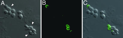
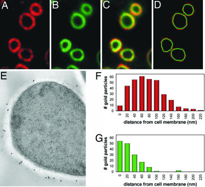
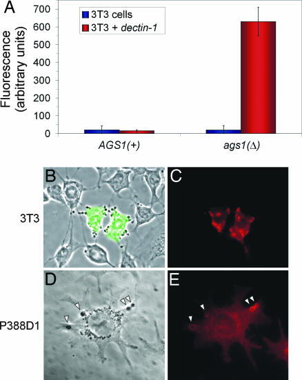
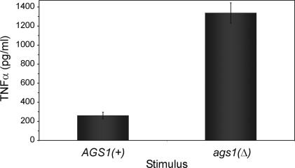
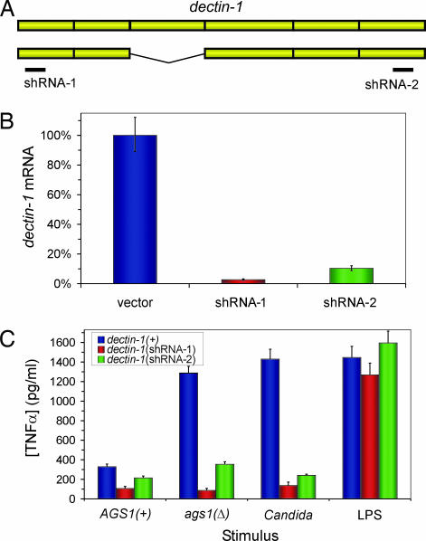
Successful infection by fungal pathogens depends on subversion of host immune mechanisms that detect conserved cell wall components such as beta-glucans. A less common polysaccharide, alpha-(1,3)-glucan, is a cell wall constituent of most fungal respiratory pathogens and has been correlated with pathogenicity or linked directly to virulence. However, the precise mechanism by which alpha-(1,3)-glucan promotes fungal virulence is unknown. Here, we show that alpha-(1,3)-glucan is present in the outermost layer of the Histoplasma capsulatum yeast cell wall and contributes to pathogenesis by concealing immunostimulatory beta-glucans from detection by host phagocytic cells. Production of proinflammatory TNFalpha by phagocytes was suppressed either by the presence of the alpha-(1,3)-glucan layer on yeast cells or by RNA interference based depletion of the host beta-glucan receptor dectin-1. Thus, we have functionally defined key molecular components influencing the initial host-pathogen interaction in histoplasmosis and have revealed an important mechanism by which H. capsulatum thwarts the host immune system. Furthermore, we propose that the degree of this evasion contributes to the difference in pathogenic potential between dimorphic fungal pathogens and opportunistic fungi.


Conflict of interest statement

The authors declare no conflict of interest.

Figures

Fig. 1.
Fig. 1.
α-(1,3)-Glucan is produced during the transition from conidia to yeast. Histoplasma conidia were incubated for 18 h at 37°C in F-12 medium containing 10% FBS and visualized by direct interference contrast microscopy (DIC) (A) and immunofluorescence microscopy (B) after staining with an antibody specific for α-(1,3)-glucan. Arrowheads indicate germinating conidia with emergent yeast buds that have a smoother cell surface. (C) Merged DIC and immunofluorescence images show that α-(1,3)-glucan is present only around the yeast buds and not the conidial structure.
Fig. 2.
Fig. 2.
α-(1,3)-Glucan comprises the outer cell wall layer of yeast. Wild-type Histoplasma yeasts were fixed, and antibodies specific for α-(1,3)-glucan (A) and β-(1,3)-glucan (B) were used to localize the respective polysaccharides. (C) Merged α-(1,3)-glucan and β-(1,3)-glucan localizations show a somewhat layered spatial organization. (D) Tracings of the α-(1,3)- and β-(1,3)-glucan outer perimeters (red and green, respectively) were overlaid, confirming that α-(1,3)-glucan forms the outermost surface. (E) Representative immunoelectron micrograph (n = 25) in which α-(1,3)-glucans and β-glucans were labeled in wild-type yeast cells with 18-nm and 12-nm gold particles, respectively. (F and G) Histograms showing the distribution of individual α-(1,3)-glucan (F; n = 342) and β-(1,3)-glucan (G; n = 162) labeling in electron micrographs, calculated as the perpendicular distance from the cell membrane.
Fig. 3.
Fig. 3.
The α-(1,3)-glucan layer blocks recognition of Histoplasma yeast by dectin-1. (A) Live Histoplasma yeasts lacking α-(1,3)-glucan [ags1(Δ)] readily bind to dectin-1-expressing 3T3 cells, whereas yeasts possessing α-(1,3)-glucan [AGS1(+)] do not bind. Binding was measured by coincubation of Histoplasma yeast with confluent monolayers of 3T3 cells or dectin-1-expressing 3T3 cells followed by quantitation of bound yeasts with the fungal fluorescent stain Uvitex 3BSA. (B and C) Binding of ags1(Δ) yeast cells depends on dectin-1 expression. (B) Merged phase-contrast and GFP fluorescence images show that only 3T3 cells expressing dectin-1 (marked by cotransfection of a gfp expression plasmid) bind ags1(Δ) yeast. (C) Immunofluorescence labeling of cells with an antibody to dectin-1 shows enrichment of dectin-1 protein at sites of contact with Histoplasma ags1(Δ) yeast. (D and E) P388D1 macrophage-like cells were incubated with ags1(Δ) yeast (arrowheads) for 30 min, and dectin-1 localization was determined. Phase-contrast image (D) and immunofluorescent labeling with an antibody to dectin-1 (E) show enrichment of dectin-1 around Histoplasma-containing phagosomal compartments.
Fig. 4.
Fig. 4.
α-(1,3)-Glucan reduces TNFα production by phagocytic cells in response to Histoplasma yeasts. ELISA-based quantitation of TNFα produced by P388D1 phagocytic cells in response to 3-h infection by live Histoplasma yeast shows increased TNFα stimulation by yeasts that lack the α-(1,3)-glucan polysaccharide compared with yeasts surrounded by α-(1,3)-glucan. All results represent data obtained from triplicate infections, with error bars corresponding to the SD.
Fig. 5.
Fig. 5.
dectin-1 mediates the functional response to Histoplasma yeasts that lack α-(1,3)-glucan. (A) Schematic representation depicts the two major splice isoforms of dectin-1 and the location targeted for RNAi by two dectin-1 shRNAs. (B) Depletion of dectin-1 was confirmed by quantitative RT-PCR of dectin-1 mRNA in P388D1 clonal lines harboring an empty RNAi vector or shRNAs targeting dectin-1. (C) TNFα produced by P388D1 lines in response to live Histoplasma yeasts with and without the α-(1,3)-glucan cell wall layer [AGS1(+) and ags1(Δ), respectively], live C. albicans cap1/cap1 yeast (the cap1 mutation impairs filamentous growth of Candida), or 39 endotoxin units/ml of endotoxin (LPS) was quantified by ELISA. The dectin-1(+) cell line used was a stable transfectant line expressing a gfp-targeting shRNA. Results represent data obtained from three infections, and error bars indicate SD.

References

    1. Brown GD. Nat Rev Immunol. 2006;6:33–43. - PubMed
    1. Palma AS, Feizi T, Zhang Y, Stoll MS, Lawson AM, Diaz-Rodriguez E, Campanero-Rhodes MA, Costa J, Gordon S, Brown GD, et al. J Biol Chem. 2006;281:5771–5779. - PubMed
    1. Nemecek JC, Wuthrich M, Klein BS. Science. 2006;312:583–588. - PubMed
    1. Medoff G, Sacco M, Maresca B, Schlessinger D, Painter A, Kobayashi GS, Carratu L. Science. 1986;231:476–479. - PubMed
    1. Klimpel KR, Goldman WE. Infect Immun. 1988;56:2997–3000. - PMC - PubMed

Publication types