. 2007 Jan 23;104(4):1325-30.
doi: 10.1073/pnas.0605208103. Epub 2007 Jan 16.

Progressive parkinsonism in mice with respiratory-chain-deficient dopamine neurons

Affiliations

Progressive parkinsonism in mice with respiratory-chain-deficient dopamine neurons

Mats I Ekstrand et al. Proc Natl Acad Sci U S A. .

Abstract

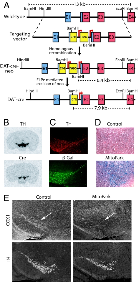
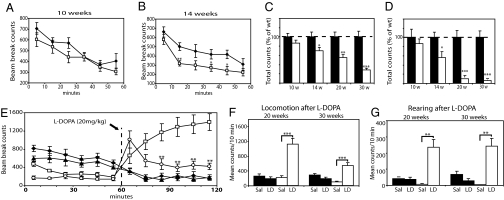
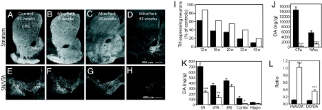
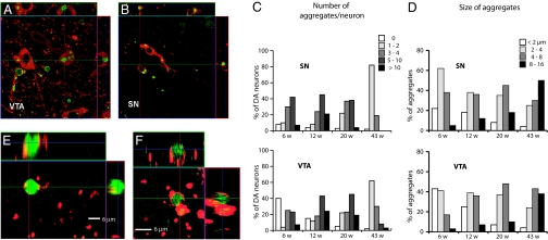
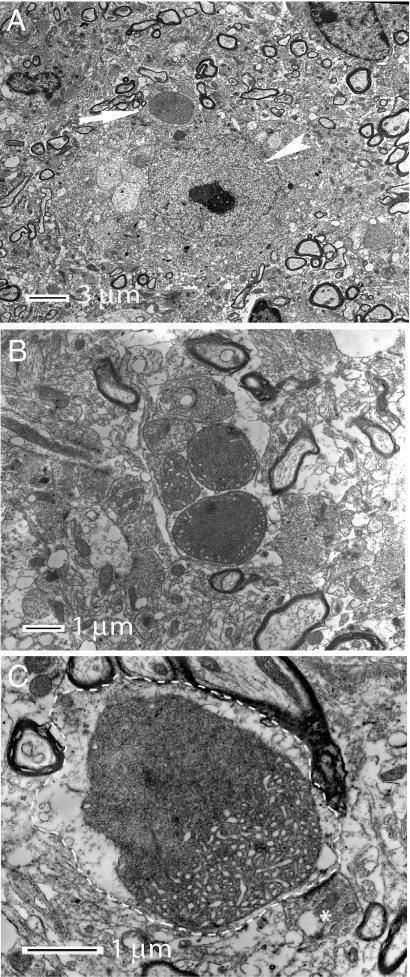
Mitochondrial dysfunction is implicated in the pathophysiology of Parkinson's disease (PD), a common age-associated neurodegenerative disease characterized by intraneuronal inclusions (Lewy bodies) and progressive degeneration of the nigrostriatal dopamine (DA) system. It has recently been demonstrated that midbrain DA neurons of PD patients and elderly humans contain high levels of somatic mtDNA mutations, which may impair respiratory chain function. However, clinical studies have not established whether the respiratory chain deficiency is a primary abnormality leading to inclusion formation and DA neuron death, or whether generalized metabolic abnormalities within the degenerating DA neurons cause secondary damage to mitochondria. We have used a reverse genetic approach to investigate this question and created conditional knockout mice (termed MitoPark mice), with disruption of the gene for mitochondrial transcription factor A (Tfam) in DA neurons. The knockout mice have reduced mtDNA expression and respiratory chain deficiency in midbrain DA neurons, which, in turn, leads to a parkinsonism phenotype with adult onset of slowly progressive impairment of motor function accompanied by formation of intraneuronal inclusions and dopamine nerve cell death. Confocal and electron microscopy show that the inclusions contain both mitochondrial protein and membrane components. These experiments demonstrate that respiratory chain dysfunction in DA neurons may be of pathophysiological importance in PD.


Conflict of interest statement

Conflict of interest statement: N.-G.L., L.O., and M.I.E. are coowners of a company owning commercial rights to the MitoPark mice.

Figures

Fig. 1.
Fig. 1.
Creation of MitoPark mice. (A) Creation of DAT-cre mice. A cassette containing an NLS-cre (cre) gene followed by an FRT-flanked (arrows) neomycin (neo) gene was inserted upstream of the translation start codon in exon 2. The neo gene was removed by breeding to FLPe deleter mice. Noncoding exon sequence is blue, and coding sequence is red. (B) In situ hybridization of midbrain dopamine neurons. Probes detecting TH and cre mRNA showed overlapping hybridization signals in SN and VTA of a heterozygous DAT-cre mouse. (C) Immunohistochemistry of a ROSA26-R reporter mouse containing the DAT-cre knock-in allele. The cre protein expression activates β-galactosidase protein expression in a pattern that overlaps with TH protein expression in SN and VTA. (D) Cytochrome c oxidase (COX) activity in a 20-week-old control mouse is normal in VTA, whereas there is dramatic decrease in COX activity (blue staining represents COX deficient neurons) in a MitoPark mouse of the same age. (E) In situ hybridization shows decreased expression of the cytochrome c oxidase subunit I (COX I) mRNA in DA neurons of the midbrain (arrow) of a 6-week-old MitoPark mouse (Upper Right) in comparison with a control (Upper Left). In situ hybridization was performed to detect TH mRNA expression in DA neurons of SN and VTA in adjacent sections (Lower).
Fig. 2.
Fig. 2.
Analysis of spontaneous activity and motor function in MitoPark mice. (A–D) Locomotion and rearing of MitoPark (white dots/bars; n = 8) and control mice (black dots/bars; n = 7) were measured for 60 min in open-field activity cages. Locomotion in 10-week-old MitoPark mice was not significantly different from age-matched controls (A), whereas 14-week-old MitoPark mice displayed decreased locomotion (B). Total locomotion (C) and total rearing (D) during 60 min decreased with increasing age in MitoPark mice (n = 12), but not in age-matched controls (n = 12). Error bars indicate ± SEM. Statistically significant differences to age-matched controls are indicated as: ∗, P < 0.05; ∗∗, P < 0.01; ∗∗∗, P < 0.001. (E–G) Cohorts of 20- and 30-week-old MitoPark (white dots/bars; n = 12) and control mice (black dots/bars; n = 15) were treated with L-DOPA (20 mg/kg) or saline. (E) Locomotion in controls at age 20 weeks (black squares), controls at age 30 weeks (black triangles), MitoPark mice at age 20 weeks (white squares), and MitoPark mice at age 30 weeks (white circles). MitoPark mice of both ages responded to L-DOPA treatment with increased locomotion (E and F) and rearing (G). L-DOPA treatment of younger MitoPark mice resulted in a greater locomotion response than treatment of older MitoPark mice (E). L-DOPA has no effect in control mice (E and F). The increase of rearing after L-DOPA treatment of MitoPark mice was similar in both age groups (G). Mean ± SEM is indicated by bars. The bars show mean locomotion (F) and mean rearing (G) in control and MitoPark mice treated with saline (Sal) or L-DOPA (LD). Statistically significant differences are indicated: ∗, P < 0.05; ∗∗, P < 0.01; ∗∗∗, P < 0.001.
Fig. 3.
Fig. 3.
TH immunohistochemistry and DA levels in the midbrain of MitoPark mice. (A–D) TH immunoreactivity in striatum of control (A) and MitoPark mice (B–D). (E–H) TH immunoreactivity in SN and VTA of control (E) and MitoPark (F and G) mice. (I) Quantitative assessment of cell loss in SN (black bars) and VTA (white bars) of MitoPark mice at different ages. (J and K) Measurement of DA and DA metabolite levels. DA levels in striatum (J) and other brain regions (K) of 20-week-old control (black bars) and MitoPark (white bars) mice. EM, eminentia mediana (=A12 area), hippo: the hippocampal formation. (L) Ratios of the DA metabolites HVA and DOPAC (DO) to DA in striatum of 20-week-old control (black bars) and MitoPark (white bars) mice. Error bars indicate ± SEM. Statistically significant differences to age-matched controls are indicated as: ∗, P < 0.05; ∗∗, P < 0.01; ∗∗∗, P < 0.001.
Fig. 4.
Fig. 4.
Analysis of intracellular inclusions. (A and B) Confocal microscopy images of immunoreactive inclusions (green) in TH immunoreactive DA neurons (red) of VTA and SN. (C) Quantification of number of aggregates per DA neuron in SN and VTA of MitoPark mice at different ages. (D) Size of aggregates in DA neurons in SN and VTA of MitoPark mice at different ages. (E and F) High magnification confocal images of aggregates after double immunofluorescence labeling to detect inclusions (green) and mitochondria (red).
Fig. 5.
Fig. 5.
Electron micrographs illustrating the morphology of intraneuronal inclusions. (A) An inclusion (arrow) in a nerve cell with nucleolus-containing nucleus (arrowhead). (B) A cluster of five abnormal mitochondria and inclusions containing mitochondrial membranes. (C) High magnification of a single inclusion of mixed morphology, which is located in a dendrite (delineated by dashed line). The amorphous portion of the inclusion shows diffuse lining and dissolved membrane whereas the more intact portion of the body show tubular structures and a visible double layer membrane. A bouton making synaptic contact with the dendrite is indicated by an asterisk.

References

    1. Gaspari M, Larsson NG, Gustafsson CM. Biochim Biophys Acta. 2004;1659:148–152. - PubMed
    1. Dauer W, Przedborski S. Neuron. 2003;39:889–909. - PubMed
    1. Shen J, Cookson MR. Neuron. 2004;43:301–304. - PubMed
    1. Park J, Lee SB, Lee S, Kim Y, Song S, Kim S, Bae E, Kim J, Shong M, Kim JM, Chung J. Nature. 2006;441:1157–1161. - PubMed
    1. Clark IE, Dodson MW, Jiang C, Cao JH, Huh JR, Seol JH, Yoo SJ, Hay BA, Guo M. Nature. 2006;441:1162–1166. - PubMed

Publication types