. 2007 Jan 15;176(2):173-82.
doi: 10.1083/jcb.200607073.

Cell cycle progression and de novo centriole assembly after centrosomal removal in untransformed human cells

Affiliations

Cell cycle progression and de novo centriole assembly after centrosomal removal in untransformed human cells

Yumi Uetake et al. J Cell Biol. .

Abstract

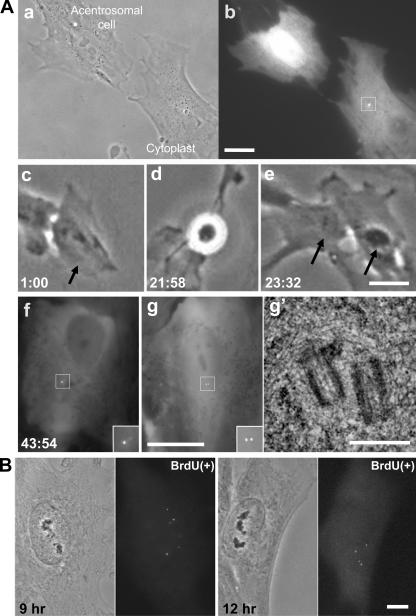
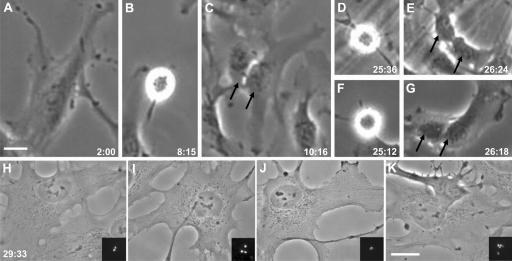
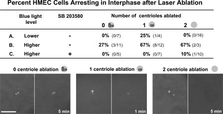
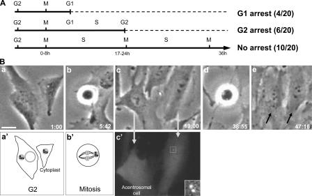
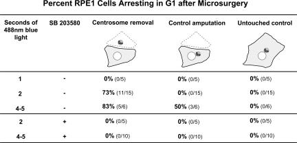
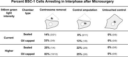
How centrosome removal or perturbations of centrosomal proteins leads to G1 arrest in untransformed mammalian cells has been a mystery. We use microsurgery and laser ablation to remove the centrosome from two types of normal human cells. First, we find that the cells assemble centrioles de novo after centrosome removal; thus, this phenomenon is not restricted to transformed cells. Second, normal cells can progress through G1 in its entirety without centrioles. Therefore, the centrosome is not a necessary, integral part of the mechanisms that drive the cell cycle through G1 into S phase. Third, we provide evidence that centrosome loss is, functionally, a stress that can act additively with other stresses to arrest cells in G1 in a p38-dependent fashion.


Figures

Figure 1.
Figure 1.
De novo centriole assembly in an acentrosomal RPE1 cell. (A, a) Acentrosomal cell and cytoplast 30 min after G1 microsurgery. (b) Fluorescence image of the GFP-tagged centriole pair (box) in the cytoplast. (c) The same acentrosomal cell at 1 h after microsurgery (arrow). (d and e) This acentrosomal cell enters mitosis and divides into two daughters (arrows). (f) Fluorescence images of one daughter showing bright focal centrin-1/GFP spots (inset). The inset shows the centrin foci at higher magnification. (g) The other daughter cell that was serial-sectioned for EM analysis. (g′) Electron micrograph of a section showing two centrioles in the location of the fluorescent dots. Hours:minutes after the microsurgery are shown in the lower corner of each frame taken from the time-lapse recording. (B) Paired phase and fluorescence images of two cells 9 and 12 h after G1 centrosome removal. These cells are in S phase, as determined by BrdU incorporation. The fluorescence images show the several centrin-1/GFP foci (precentrioles) that have assembled. Bars: (A, a–g) 20 μm; (A, g′) 0.5 μm; (B) 10 μm.
Figure 2.
Figure 2.
Acentrosomal RPE1 cell progressing from G1 through two mitoses. (A) Acentrosomal cell 2 h after the microsurgery. (B) First mitosis. (C) Progeny of the first mitosis. The two daughters are indicated by arrows. (D and E and F and G) Second mitosis of both daughters. (H–K) The four granddaughters are shown at higher magnification, with insets showing the variable number of centrin-1/GFP foci that are indicative of centrioles assembled de novo. Phase-contrast images with GFP fluorescence (z series, maximum intensity point projections) shown in the insets. Hours:minutes after the microsurgery are shown in the bottom corner of each frame taken from the time-lapse recording. Bars, 20 μm.
Figure 3.
Figure 3.
Laser ablation of one or two centrioles during G1 predisposes HMEC cells to a p38-dependent interphase arrest. (top) The blue light level column shows the intensity of the confocal blue light power used to position cells at the coordinates of the laser beam and, later, follow the cells for 72 h. Lower, 107 μW output at the objective; higher, 450 μW output at the objective. The SB 203580 column shows the presence (+) or absence (−) of the p38 inhibitor. In the number of centrioles ablated columns, a near miss did not ablate any centrioles (0) and these serve as controls; other ablations eliminated one centriole (1), and others eliminated both centrioles (2). Line A summarizes the results for cells positioned in the coordinates of the laser beam and later followed for 72 h at the lower 488-nm blue light level in the absence of the p38 inhibitor SB 203580. Line B shows the results for cells positioned in the coordinates of the laser beam and, later, followed at the higher blue light level without p38 inhibitor. Line C shows the results for cells exposed to the higher blue light level in the presence of SB 203580 starting 30 min before laser ablation and followed thereafter in the presence of the inhibitor. (bottom) Before and after fluorescence images of 0, 1, and 2 centriole laser ablations. Times in the right hand panels indicate minutes after ablation. Bar, 5 μm.
Figure 4.
Figure 4.
RPE1 cells “born” without centrosomes progress through G1 in its entirety. (A) Summary of data. G2 cells were identified by the presence of four centrin dots (two centrosomes) and were cut to remove one centrosome. They were then followed by phase-contrast time-lapse video microscopy for at least 36 h. After first mitosis, BrdU was added to the medium and acentriolar daughters were identified. Those acentrosomal daughters that failed to enter second mitosis were fixed to assay for BrdU incorporation. First and second mitoses occurred between 0–8 h and 17–24 h, respectively, after the microsurgery. (B) Cell cycle progression of a cell from which one centrosome was removed in G2. (a and a′) Image and diagram of cell/cytoplast 1 h after microsurgery. (b and b′) Image and diagram of cell with one centrosome in mitosis. (c and c′) Phase and centrin-1/GFP fluorescence images of the daughter cells. The cell on the left has inherited no centrioles, and the one on the right has inherited the centriole pair (box and inset at higher magnification; z series maximum intensity projection). The acentrosomal cell enters mitosis (d) and produces two daughters, indicated by arrows (e). Hours:minutes after microsurgery are shown in the lower corners of each frame taken from time-lapse recording. Bar, 20 μm.
Figure 5.
Figure 5.
Microsurgery and centrosome removal predispose RPE1 cells to p38-dependent G1 arrest. For each experiment, a G1 cell was microsurgically cut to remove the centrosome and, in the same field, a cell was cut to amputate an equivalent portion of the cytoplasm without removing the centrosome. Untouched cells in the same field served as controls. BrdU was added to allow for later analysis for entry into S phase. 30 min after the microsurgery, the field was exposed for defined durations to the 488-nm light. Each field was followed by phase-contrast time-lapse microscopy for at least 48 h to determine if the acentrosomal cell, control amputation, or untouched controls progressed to mitosis in the presence of BrdU. The acentrosomal cells and control cut cells that arrested in interphase were fixed and assayed for BrdU incorporation; none showed any BrdU incorporation, demonstrating that the cells never progressed out of G1. The last two lines of this table show the results of experiments in which the preparations continuously contained the p38 stress kinase inhibitor SB 203580 starting 30 min before the microsurgery.
Figure 6.
Figure 6.
G1 progression of acentrosomal and control cut BSC-1 cells under various experimental conditions. For each experiment, an interphase cell was microsurgically cut to remove the centrosome and, in the same field, a cell was cut to amputate an equivalent portion of the cytoplasm without removing the centrosome. Untouched cells in the same field served as controls. After microsurgery in oil-capped micromanipulation chambers, cells were continuously cultured in the same chamber or transferred into a sealed filming chamber, and observed for ∼90 h at the indicated intensity of 546-nm green light. After microsurgery, all cells went through mitosis, but thereafter some arrested in interphase. The Green light level column shows green light intensity used for time-lapse imaging. Current, 4.75 nW condenser output; Higher, 1170 nW condenser output. In the Chamber type column shows the chamber types used. Sealed, the sealed chamber currently used for time-lapse observation; Oil capped, the oil-capped chamber used for micromanipulation.

References

    1. Ambrosino, C., and A.R. Nebreda. 2001. Cell cycle regulation by p38 MAP kinases. Biol. Cell. 93:47–51. - PubMed
    1. Bohmer, R.M., E. Scharf, and R.K. Assoian. 1996. Cytoskeletal integrity is required throughout the mitogen stimulation phase of the cell cycle and mediates the anchorage-dependent expression of cyclin D1. Mol. Biol. Cell. 7:101–111. - PMC - PubMed
    1. Doxsey, S., D. McCollum, and W. Theurkauf. 2005. a. Centrosomes in cellular regulation. Annu. Rev. Cell Dev. Biol. 21:411–434. - PubMed
    1. Doxsey, S., W. Zimmerman, and K. Mikule. 2005. b. Centrosome control of the cell cycle. Trends Cell Biol. 15:303–311. - PubMed
    1. Ehrhardt, A.G., and G. Sluder. 2005. Spindle pole fragmentation due to proteasome inhibition. J. Cell. Physiol. 204:808–818. - PubMed

Publication types

MeSH terms