. 2007 Jan 15;176(2):223-30.
doi: 10.1083/jcb.200606116.

Involvement of zebrafish Na+,K+ ATPase in myocardial cell junction maintenance

Affiliations

Involvement of zebrafish Na+,K+ ATPase in myocardial cell junction maintenance

Elena Cibrián-Uhalte et al. J Cell Biol. .

Abstract

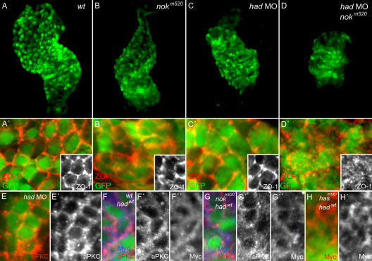
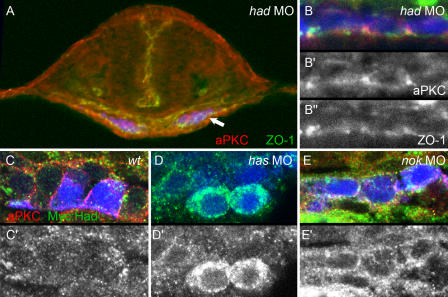
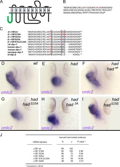
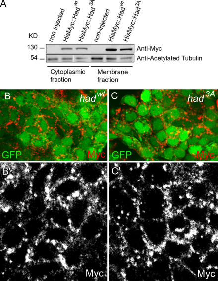
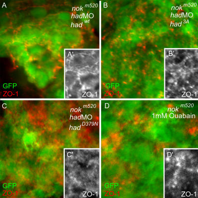
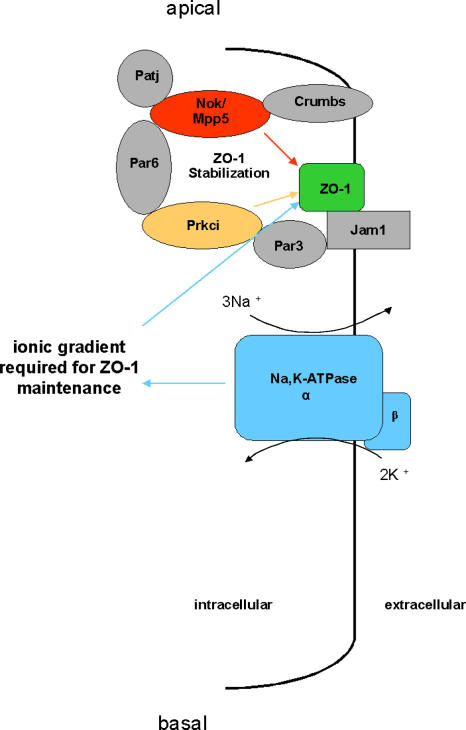
Na(+),K(+) ATPase is an essential ion pump involved in regulating ionic concentrations within epithelial cells. The zebrafish heart and mind (had) mutation, which disrupts the alpha1B1 subunit of Na(+),K(+) ATPase, causes heart tube elongation defects and other developmental abnormalities that are reminiscent of several epithelial cell polarity mutants, including nagie oko (nok). We demonstrate genetic interactions between had and nok in maintaining Zonula occludens-1 (ZO-1)-positive junction belts within myocardial cells. Functional tests and pharmacological inhibition experiments demonstrate that Na(+),K(+) ATPase activity is positively regulated via an N-terminal phosphorylation site that is necessary for correct heart morphogenesis to occur, and that maintenance of ZO-1 junction belts requires ion pump activity. These findings suggest that the correct ionic balance of myocardial cells is essential for the maintenance of epithelial integrity during heart morphogenesis.


Figures

Figure 1.
Figure 1.
Genetic interactions of had and nok during heart morphogenesis. Reconstructions of confocal z-stack sections of embryonic hearts. (A–D) Morphology of transgenic Tg(cmlc2:GFP) embryonic hearts in different genetic backgrounds at 34–36 hpf. (A′–D′) Localization of ZO-1 in myocardial cells at 36 hpf in different genetic backgrounds and details thereof (insets). Whereas ZO-1–positive junction belts are present in nokm520 mutants (B and B′) and had morphants (C and C′), they are severely disrupted upon loss of both genes (D and D′). (E and E′) aPKCs are correctly localized to the membrane in had morphants at 34–36 hpf. (F–H) Embryos of different genetic backgrounds injected with mRNA encoding Myc-tagged Had/Na+,K+ ATPase were used to detect the subcellular localization of the fusion protein, which remains at the membrane in wt (F), nokm520 (G), and hasm567 mutants (H).
Figure 2.
Figure 2.
Effects of had, has, and nok morphants on myocardial apical–basal polarity at the 20-somite stage. Transverse sections of heart cone stage (20-somite) embryos. GFP is false-colored in blue; aPKC, red (A–C and E) or gray (B′); Myc∷Had, green (C–E) or gray (C′–E′); ZO-1, green (A and B) or gray (B″). (A) had morphant with a section plane through the middle of the heart cone. The two bilateral wings of myocardial cells are blue. Arrow indicates the lateral portion of the myocardial field that was used for detail images within the various genetic backgrounds. (B, B′, and B″) had morphants are correctly polarized and display apical ZO-1– and aPKC-positive spots. (C and C′) In wt myocardial cells, levels of Myc∷Had/Na+,K+ ATPase are low, and a clear localization pattern is not apparent. (D and D′) has morphant myocardial cells exhibit higher levels of Myc∷Had/Na+,K+ ATPase, which is localized around the circumference of cells. (E and E′) Similarly, nok morphants display localization of Myc∷Had/Na+,K+ ATPase and aPKC around the entire myocardial circumferences, which is indicative of loss of apical–basal polarity within these cells.
Figure 3.
Figure 3.
N-terminal Ser25 is required for correct activity of Had/Na+,K+ ATPase during heart tube elongation. (A) Predicted structure of the Na+,K+ ATPase α1B1 subunit. The N-terminal intracellular tail (98 residues) is shown in green. Transmembrane domains are numbered. (B) Protein sequence of the Had/Na+,K+ ATPase N-terminal intracellular tail (first 98 residues). Three predicted aPKC phosphorylation target sites are shown in red. (C) Sequence alignment of the first 35 residues of the N-terminal intracellular tail of zebrafish wt and mutated forms of Na+,K+ ATPase α1B1, rat Na+,K+ ATPase α1, human Na+,K+ ATPase α1, mouse Na+,K+ ATPase α1, X. laevis Na+,K+ ATPase α1, and zebrafish Na+,K+ ATPase α2. Predicted aPKC phosphorylation target sites Ser16, which is conserved among all species, Ser23, which is conserved among rat and zebrafish, and Ser25, which is only present in zebrafish, are shown in red. Serine residues mutated to alanine are shown in blue. (D–I) Dorsal view of embryos at 34–36 hpf analyzed for expression of myocardial marker cmlc2. Heart tube elongation defects in hadla1 mutant embryos that were injected with wt or mutant forms of had mRNA. (J) Quantifications of the rescue of hadla1 mutant heart tube defects after injection of wt or mutant forms of had mRNA. Statistical P values for rescue efficiency of different mutant mRNAs compared with wt mRNA rescue efficiency are shown. P values <0.05 are considered statistically significant.
Figure 4.
Figure 4.
The N-terminal phosphorylation state of Had/Na+,K+ ATPase does not affect membrane association. (A) Western blot of 32 hpf embryonic extracts shows that HisMyc∷Hadwt and HisMyc∷Had3A are enriched within the membrane and cytoskeletal fraction. (B and C) Confocal fluorescence microscopic images. Myocardial wt cells expressing HisMyc∷Hadwt (B and B′) and HisMyc∷Had3A (C and C′). Both recombinant forms of the Na pump are mostly localized at the outer cell membrane.
Figure 5.
Figure 5.
Genetic interactions of a Had/Na+,K+ ATPase regulatory and catalytic mutants with Nok/Mpp5 in the maintenance of ZO-1–positive junction belts. Reconstructions of confocal z-stack sections of embryonic hearts. (A–D) nokm520 mutants transgenic for Tg(cmlc2:GFP) were injected with hadMO together with HisMyc∷hadwt (A), HisMyc∷had3A (B), HisMyc∷hadD379N mRNA (C), or treated with 1 mM ouabain (D). HisMyc∷hadwt mRNA rescues the disruption of ZO-1–positive junction belts in nokm520 mutants (A′), unlike HisMyc∷had3A (B′) and HisMyc∷hadD379N mutant mRNA, which does not (C′), or ouabain treatment (D′).
Figure 6.
Figure 6.
Model for the interactions between Nok/Mpp5 and Had/Na+,K+ ATPase in the maintenance of apical junction belts within myocardial cells. The ion pump activity of Had/Na+,K+ ATPase, which maintains the ionic balance of myocardial cells, interacts with the tight junction–associated protein Nok/Mpp5, a scaffolding partner of apical Crumbs and Par6–aPKCι protein complexes, in the stabilization of apical ZO-1–positive junctional belts.

References

    1. Anzenberger, U., N. Bit-Avragim, S. Rohr, F. Rudolph, B. Dehmel, P. Haas, D. Gilmour, T.E. Willnow, and S. Abdelilah-Seyfried. 2006. Elucidation of Megalin/LRP2-dependent endocytic transport processes in the larval zebrafish pronephros. J. Cell Sci. 119:2127–2137. - PubMed
    1. Bertorello, A.M., A. Aperia, S.I. Walaas, A.C. Nairn, and P. Greengard. 1991. Phosphorylation of the catalytic subunit of Na+,K+ ATPase inhibits the activity of the enzyme. Proc. Natl. Acad. Sci. USA. 88:11359–11362. - PMC - PubMed
    1. Blanco, G., and R.W. Mercer. 1998. Isozymes of the Na+,K+ ATPase: heterogeneity in structure, diversity in function. Am. J. Physiol. 275:F633–F650. - PubMed
    1. Chen, J., and M. Fishman. 1996. Zebrafish tinman homolog demarcates the heart field and initiates myocardial differentiation. Development. 122:3809–3816. - PubMed
    1. Chibalin, A.V., A.I. Katz, P.O. Berggren, and A.M. Bertorello. 1997. Receptor-mediated inhibition of renal Na+,K+ ATPase is associated with endocytosis of its alpha- and beta-subunits. Am. J. Physiol. 273:C1458–C1465. - PubMed

Publication types

MeSH terms