. 2007 Jan 22;204(1):129-39.
doi: 10.1084/jem.20061115. Epub 2007 Jan 16.

Hematopoietic reconstitution by multipotent adult progenitor cells: precursors to long-term hematopoietic stem cells

Affiliations

Hematopoietic reconstitution by multipotent adult progenitor cells: precursors to long-term hematopoietic stem cells

Marta Serafini et al. J Exp Med. .

Erratum in

  • J Exp Med. 2007 Jul 9;204(7):1729

Abstract

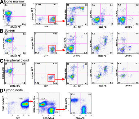
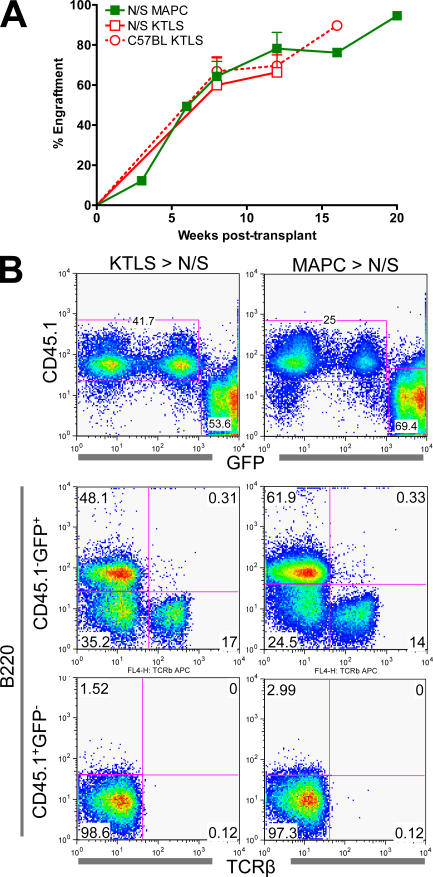
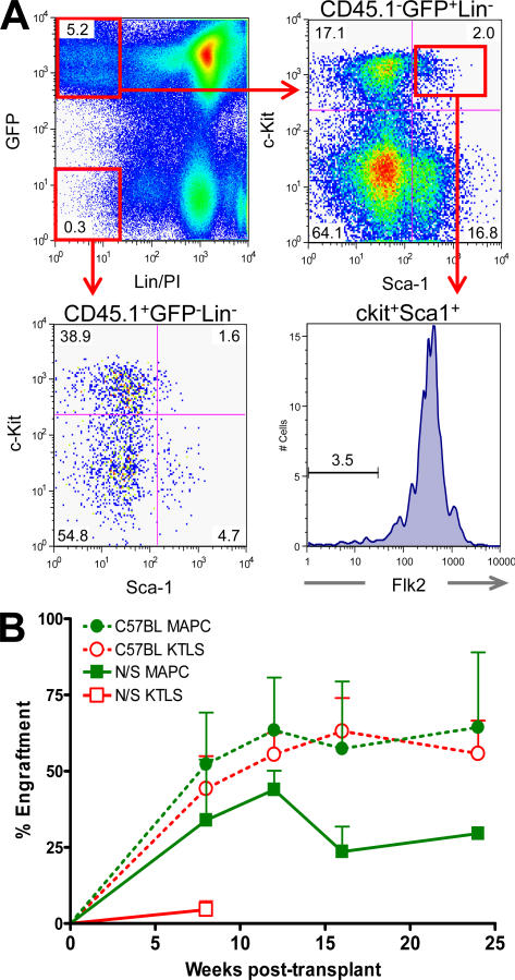
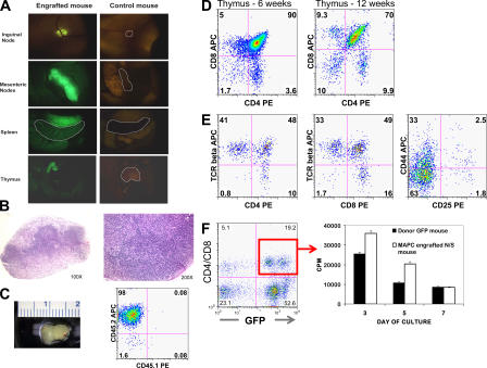
For decades, in vitro expansion of transplantable hematopoietic stem cells (HSCs) has been an elusive goal. Here, we demonstrate that multipotent adult progenitor cells (MAPCs), isolated from green fluorescent protein (GFP)-transgenic mice and expanded in vitro for >40-80 population doublings, are capable of multilineage hematopoietic engraftment of immunodeficient mice. Among MAPC-derived GFP+CD45.2+ cells in the bone marrow of engrafted mice, HSCs were present that could radioprotect and reconstitute multilineage hematopoiesis in secondary and tertiary recipients, as well as myeloid and lymphoid hematopoietic progenitor subsets and functional GFP+ MAPC-derived lymphocytes that were functional. Although hematopoietic contribution by MAPCs was comparable to control KTLS HSCs, approximately 10(3)-fold more MAPCs were required for efficient engraftment. Because GFP+ host-derived CD45.1+ cells were not observed, fusion is not likely to account for the generation of HSCs by MAPCs.


Figures

Figure 1.
Figure 1.
Hematopoietic reconstitution from MAPCs grafted in NOD-SCID mice. 106 GFP+CD45.2+ MAPCs were transplanted in sublethally irradiated NOD-SCID mice treated with anti–asialo-GM1 antibodies 4 h before transplantation and on days +11 and +22. Representative flow cytometry profiles of BM, spleen, PB, and LNs of NOD-SCID (CD45.1+) mice ≥12 wk after transplant demonstrating multilineage (B lymphoid, T lymphoid, and myeloid cells) reconstitution. Contour plots shown are after gating on GFP fraction, as indicated.
Figure 2.
Figure 2.
Hematopoietic reconstitution from KTLS HSCs versus MAPCs. 2–3 × 10⁵ Sca-1–depleted, host-derived BM cells were co-transplanted with 600 KTLS or 0.75 × 10⁶ MAPCs from eGFP+CD45.2+ donor mice into sublethally irradiated NOD-SCID (N/S) or C57BL-CD45.1+ recipients. (A) Mice were evaluated at intermittent time points for PB eGFP+CD45.2+ cells. (B) Representative multilineage reconstitution of KTLS- versus MAPC-engrafted NOD-SCID mice at week 12.
Figure 3.
Figure 3.
MAPCs generate KLS Flk2 HSCs and engraft secondary recipients. 0.75 × 10⁶ eGFP MAPCs were transplanted into lethally irradiated NOD-SCID mice with 2 × 10⁵ Sca-1–depleted cells of host origin. 20 wk after transplant, 106 BM cells were serially transplanted into lethally irradiated NOD-SCID or C57BL-CD45.1+ hosts. (A) 20 wk after transplant, MAPC-engrafted animals were killed and BM was evaluated for the presence of eGFP+CD45.2+ hematopoietic stem and progenitor cells. (B) PB was obtained periodically to assess primary donor (eGFP+CD45.2+) contribution and multilineage (T cell, B cell, and myeloid) engraftment.
Figure 4.
Figure 4.
Functional lymphoid reconstitution in MAPC-engrafted animals. Multiparameter analysis was performed to determine the degree of lymphohematopoietic engraftment. (A) Intravital microscopy was performed, demonstrating GFP cells in all lymphoid organs, including the thymus, spleen, and LNs of a NOD-SCID mouse 13 wk after transplantation of 106 MAPCs. (B) Hematoxylin and eosin–stained sections of a mesenteric LNs harvested from an MAPC-engrafted NOD-SCID mouse, demonstrating normal primary lymphoid follicles. (C) Representative photograph of the thymus from a NOD-SCID mouse 12 wk after transplant with flow cytometry, demonstrating >95% GFP+CD45.2+ donor-derived cells in the thymus. Representative thymic engraftment by GFP+ MAPC-derived cells (D) 6 and 12 wk after transplant followed by (E) TCR-β, CD44, and CD25 expression profiles 12 wk after transplant. (F) At 13 wk, GFP+ CD4/CD8+ cells were isolated by FACS (95.8% purity) from the spleen of an MAPC-engrafted mouse. After stimulation by anti-CD3/CD28–coated beads, proliferation was assessed by [3H]thymidine incorporation. Control cells were harvested from the spleen of an MAPC donor background CD45.2+GFP+ animal.
Figure 5.
Figure 5.
MAPC-engrafted mouse with chronic GVHD. Intravital microscopy and histopathological findings in one NOD-SCID mouse that received 0.6 × 10⁶ MAPCs 16 wk earlier are consistent with the development of chronic GVHD. Left panels show the presence of GFP+ cells in the GVHD target organs, specifically skin (underlayer), liver, lung, and ileum. All intravital images were taken at a zoom factor of 8× and a transfer lens of 0.63× with an MZFLIII stereomicroscope (300 millisecond exposure). Right panels show the corresponding hematoxylin and eosin stains of cryosections taken from the same mouse (bar, 900 μm). The skin shows inflammation in the dermis and subdermis (black arrow), extensive epidermal hyperplasia (white arrow), and dyskeratosis (asterisk). GVHD score, 3.5 (0–4 scale). The liver has moderate inflammation around the portal triads with evidence of bile duct degeneration (black arrow). GVHD score, 3.0. The lung has peribronchiolar inflammation (b, bronchiole) and extensive parenchymal inflammation in surrounding areas with organizing alveolitis and fibrosis. GVHD score, 4.0. The ileum shows only mild inflammation of the lamina propria as is typical for chronic GVHD. GVHD score, 0.5.

Similar articles

Cited by

References

    1. Morrison, S.J., and I.L. Weissman. 1994. The long-term repopulating subset of hematopoietic stem cells is deterministic and isolatable by phenotype. Immunity. 1:661–673. - PubMed
    1. Ikuta, K., and I.L. Weissman. 1992. Evidence that hematopoietic stem cells express mouse c-kit but do not depend on steel factor for their generation. Proc. Natl. Acad. Sci. USA. 89:1502–1506. - PMC - PubMed
    1. Sorrentino, B.P. 2004. Clinical strategies for expansion of haematopoietic stem cells. Nat. Rev. Immunol. 4:878–888. - PubMed
    1. Kyba, M., R.C. Perlingeiro, and G.Q. Daley. 2002. HoxB4 confers definitive lymphoid-myeloid engraftment potential on embryonic stem cell and yolk sac hematopoietic progenitors. Cell. 109:29–37. - PubMed
    1. Wang, L., P. Menendez, F. Shojaei, L. Li, F. Mazurier, J.E. Dick, C. Cerdan, K. Levac, and M. Bhatia. 2005. Generation of hematopoietic repopulating cells from human embryonic stem cells independent of ectopic HOXB4 expression. J. Exp. Med. 201:1603–1614. - PMC - PubMed

Publication types

Substances