. 2007 Jan 22;204(1):41-7.
doi: 10.1084/jem.20062259. Epub 2007 Jan 16.

T cell self-reactivity forms a cytokine milieu for spontaneous development of IL-17+ Th cells that cause autoimmune arthritis

Affiliations

T cell self-reactivity forms a cytokine milieu for spontaneous development of IL-17+ Th cells that cause autoimmune arthritis

Keiji Hirota et al. J Exp Med. .

Abstract

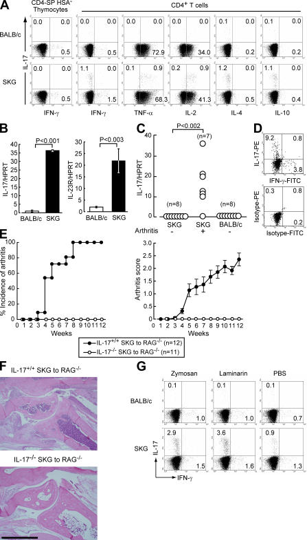
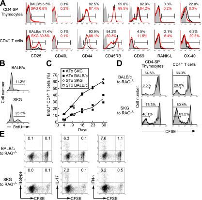
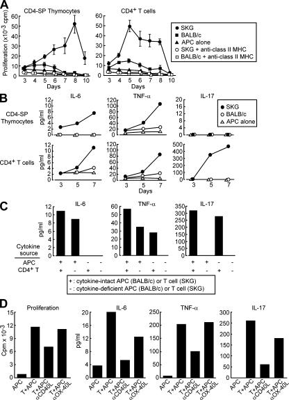
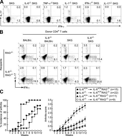
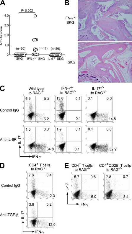
This report shows that highly self-reactive T cells produced in mice as a result of genetically altered thymic T cell selection spontaneously differentiate into interleukin (IL)-17-secreting CD4+ helper T (Th) cells (Th17 cells), which mediate an autoimmune arthritis that clinically and immunologically resembles rheumatoid arthritis (RA). The thymus-produced self-reactive T cells, which become activated in the periphery via recognition of major histocompatibility complex/self-peptide complexes, stimulate antigen-presenting cells (APCs) to secrete IL-6. APC-derived IL-6, together with T cell-derived IL-6, drives naive self-reactive T cells to differentiate into arthritogenic Th17 cells. Deficiency of either IL-17 or IL-6 completely inhibits arthritis development, whereas interferon (IFN)-gamma deficiency exacerbates it. The generation, differentiation, and persistence of arthritogenic Th17 cells per se are, however, insufficient for producing overt autoimmune arthritis. Yet overt disease is precipitated by further expansion and activation of autoimmune Th17 cells, for example, via IFN-gamma deficiency, homeostatic proliferation, or stimulation of innate immunity by microbial products. Thus, a genetically determined T cell self-reactivity forms a cytokine milieu that facilitates preferential differentiation of self-reactive T cells into Th17 cells. Extrinsic or intrinsic stimuli further expand these cells, thereby triggering autoimmune disease. Intervention in these events at cellular and molecular levels is useful to treat and prevent autoimmune disease, in particular RA.


Figures

Figure 1.
Figure 1.
Spontaneous development of arthritogenic Th17 cells in SKG mice. (A) HSA CD4-SP thymocytes or LN CD4+ T cells were stained for intracellular cytokines. (B) Quantitative RT-PCR for IL-17 and IL-23R mRNA in CD4+ T cells. Data are shown as the mean ± SD of three independent experiments. (C) Total RNA extracted from the ankle joints of individual mice with or without arthritis was subjected to quantitative RT-PCR for IL-17 mRNA. (D) CD4+ T cells infiltrating arthritic joints were stained as in A. (E) 106 CD4+ T cells from IL-17+/+ or IL-17−/− SKG mice were transferred to RAG2−/− mice. Incidence and severity of arthritis were scored every week as described previously (reference 2). Vertical bars represent the means ± SEM. (F) Histology of an ankle joint of a RAG2−/− mouse transferred with IL-17−/− or IL-17+/+ SKG CD4+ T cells (bar, 1 mm; hematoxylin and eosin staining). (G) Mice received a single i.p. injection of 2 mg zymosan or 30 mg laminarin. LN CD4+ T cells were stained for intracellular IL-17 and IFN-γ 2 wk later. Results in A, D, and G represent three to five independent experiments.
Figure 2.
Figure 2.
T cell self-reactivity and in vivo spontaneous differentiation of Th17 cells. (A) CD4-SP thymocytes and LN CD4+ T cells from 6-wk-old BALB/c or SKG mice were stained with designated mAbs. (B) Mice were given BrdU for 9 d, and LN CD4+ T cells were stained with anti-BrdU. (C) Mice that had been thymectomized at 4 wk of age were administered with BrdU for the indicated days from 6 wk of age, and percentages of BrdU-stained cells among CD4+ T cells are shown. ATx, adult thymectomy; STx, sham thymectomy. (D and E) HSA CD4-SP thymocytes or CD4+ T cells (3 × 10⁶) were labeled by CFSE and transferred to RAG2−/− mice. 5 d later, recipient splenic CD4+ T cells were assessed for CFSE profile and intracellular IL-17 and IFN-γ. Results in A, B, D, and E represent three to five independent experiments.
Figure 3.
Figure 3.
T cell self-reactivity and in vitro cytokine production. (A) HSA CD4-SP thymocytes or CD4+ T cells were cultured with autologous APCs in the presence or absence of anti–class II MHC blocking mAb. Proliferation was measured by [3H]thymidine incorporation. Vertical bars signify SD. (B) Culture supernatants in the AMLR shown in A were collected and assessed for IL-6, TNF-α, and IL-17 production. (C) CD4+ T cells from IL-6−/−, TNF-α−/−, IL-17−/−, or cytokine-intact SKG mice were cultured with cytokine-deficient or -intact APCs, and culture supernatants were collected on day 7 for cytokine assessment as in B. (D) Anti-CD40L or anti-OX40L blocking mAb (100 μg/ml) was added to the culture, and proliferation or cytokine production was assessed as shown in A and B. Results in A–D represent three independent experiments.
Figure 4.
Figure 4.
The role of IL-6 for the development of arthritogenic Th17 cells in SKG mice. (A) LN CD4+ T cells from cytokine-deficient SKG mice were stained for intracellular IL-17 and IFN-γ. (B) 2 × 10⁶ CD4+ T cells from the indicated donor mice were transferred to IL-6−/− or intact RAG2−/− mice. Intracellular IL-17 and IFN-γ in recipient splenic CD4+ T cells were stained on day 7. (C) 106 CD4+ T cells from IL-6+/+ or IL-6−/− SKG mice were transferred to IL-6+/+ or IL-6−/− RAG2−/− mice. Incidence and severity of arthritis in four groups of mice were assessed every week. Vertical bars represent the means ± SEM of scores. In comparison of four groups (•, IL-6+/+→IL-6+/+RAG−/−; ▪, IL-6−/−→IL-6+/+RAG−/−; ▴, IL-6+/+→IL-6−/−RAG−/−; ◯, IL-6−/−→IL-6−/−RAG−/−), statistically significant (P < 0.05) differences in scores are: • versus ▪, 5–12 wk; • versus ▴, 5–12 wk; • versus ◯, 5–12 wk; ▪ versus ◯, 9–12 wk; ▴ versus ◯, 9–12 wk; ▪ versus ▴, at 12 wk. Results in A and B represent three independent experiments.
Figure 5.
Figure 5.
Spontaneous development of autoimmune arthritis in IFN-γ–deficient SKG mice and IL-6–dependent cross-regulation between Th17 and Th1 cells. (A) Arthritis score in 6-mo-old cytokine-deficient SKG mice under SPF conditions. (B) Histology of an ankle joint of a 6-mo-old SKG or IFN-γ−/− SKG mouse in A. (C) CD4+ T cells from wild-type, IFN-γ−/−, or IL-17−/− mice were transferred to RAG2−/− mice, which were i.v. injected with 1 mg anti–IL-6R mAb or control rat IgG twice (on the same day and day 3). Intracellular IL-17 and IFN-γ in recipient splenic CD4+ T cells were stained on day 7. (D) CD4+ T cells from BALB/c mice were transferred to RAG2−/− mice, which were i.v. treated with 1 mg anti–TGF-β and assessed as in C. (E) BALB/c CD4+ T cells nondepleted or depleted of CD4+CD25+ T cells were transferred to RAG2−/− mice and assessed as in C. Results in C–E represent three independent experiments.

Similar articles

Cited by

References

    1. Ulmanen, I., M. Halonen, T. Ilmarinen, and L. Peltonen. 2005. Monogenic autoimmune diseases-lessons of self-tolerance. Curr. Opin. Immunol. 17:609–615. - PubMed
    1. Sakaguchi, N., T. Takahashi, H. Hata, T. Nomura, T. Tagami, S. Yamazaki, T. Sakihama, T. Matsutani, I. Negishi, S. Nakatsuru, and S. Sakaguchi. 2003. Altered thymic T-cell selection due to a mutation of the ZAP-70 gene causes autoimmune arthritis in mice. Nature. 426:454–460. - PubMed
    1. Hata, H., N. Sakaguchi, H. Yoshitomi, Y. Iwakura, K. Sekikawa, Y. Azuma, C. Kanai, E. Moriizumi, T. Nomura, T. Nakamura, and S. Sakaguchi. 2004. Distinct contribution of IL-6, TNF-alpha, IL-1, and IL-10 to T cell-mediated spontaneous autoimmune arthritis in mice. J. Clin. Invest. 114:582–588. - PMC - PubMed
    1. Chan, A.C., M. Iwashima, C.W. Turck, and A. Weiss. 1992. ZAP-70: a 70 kd protein-tyrosine kinase that associates with the TCR zeta chain. Cell. 71:649–662. - PubMed
    1. Yoshitomi, H., N. Sakaguchi, K. Kobayashi, G.D. Brown, T. Tagami, T. Sakihama, K. Hirota, S. Tanaka, T. Nomura, I. Miki, et al. 2005. A role for fungal β-glucans and their receptor Dectin-1 in the induction of autoimmune arthritis in genetically susceptible mice. J. Exp. Med. 201:949–960. - PMC - PubMed

Publication types

MeSH terms