. 2007 Feb;129(2):145-59.
doi: 10.1085/jgp.200609639. Epub 2007 Jan 16.

The location of a closed channel gate in the GABAA receptor channel

Affiliations

The location of a closed channel gate in the GABAA receptor channel

Moez Bali et al. J Gen Physiol. 2007 Feb.

Abstract

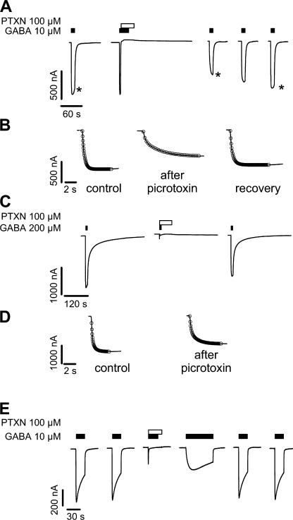
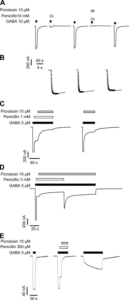
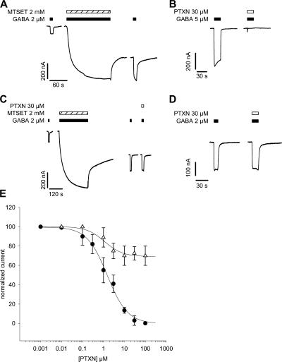
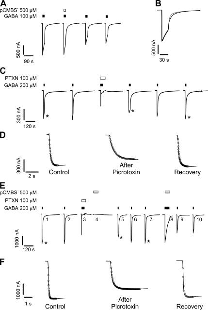
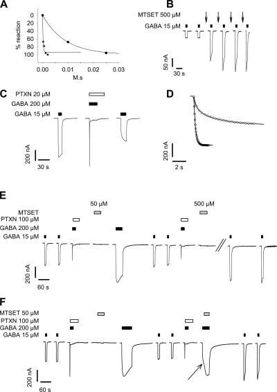
Considerable controversy surrounds the location of the closed channel gate in members of the Cys-loop receptor family of neurotransmitter-gated ion channels that includes the GABAA, glycine, acetylcholine, and 5-HT3 receptors. Cysteine-accessibility studies concluded that the gate is near the cytoplasmic end of the channel in acetylcholine and GABAA receptors but in the middle of the 5-HT3A receptor channel. Zn2+ accessibility studies in a chimeric 5-HT3-ACh receptor suggested the gate is near the channel's cytoplasmic end. In the 4-A resolution structure of the acetylcholine receptor closed state determined by cryoelectron microscopy, the narrowest region, inferred to be the gate, is in the channel's midsection from 9' to 14' but the M1-M2 loop residues at the channel's cytoplasmic end were not resolved in that structure. We used blocker trapping experiments with picrotoxin, a GABAA receptor open channel blocker, to determine whether a gate exists at a position more extracellular than the picrotoxin binding site, which is in the vicinity of alpha1Val257 (2') near the channel's cytoplasmic end. We show that picrotoxin can be trapped in the channel after removal of GABA. By using the state-dependent accessibility of engineered cysteines as reporters for the channel's structural state we infer that after GABA washout, with picrotoxin trapped in the channel, the channel appears to be in the closed state. We infer that a gate exists between the picrotoxin binding site and the channel's extracellular end, consistent with a closed channel gate in the middle of the channel. Given the homology with acetylcholine and 5-HT3 receptors there is probably a similar gate in those channels as well. This does not preclude the existence of an additional gate at a more cytoplasmic location.


Figures

Figure 1.
Figure 1.
Picrotoxin bound in the open state was trapped in the channel after channel closure and slows subsequent GABA-induced channel opening. (A) Two electrode voltage clamp recordings from wild-type α1β1γ2S expressing Xenopus oocyte. Black bars above traces indicate 10 μM GABA application; open bar, 100 μM picrotoxin (PTXN) application. In the second trace, the GABA application was begun before the coapplication of GABA and picrotoxin. The stars (*) in A indicate traces shown at expanded time scale in B. Currents during 5-min washes between traces are not shown. (B) Expanded time scale of traces from A, before (control) and after (picrotoxin) application of picrotoxin and after recovery. Exponential fit (open circles) overlays current traces (black line). In some places the black line cannot be seen because the fitted line overlaps the data completely. Note the slowed current rise time after picrotoxin application (picrotoxin) compared with before picrotoxin and after recovery. See text for average time constants. (C) Similar experiment as in A except that a saturating GABA concentration (200 μM) was used. The application of picrotoxin (open bar) is concomitant with the application of GABA. (D) Extended time scale view of the first and third traces in C. Exponential fit (open circles), currents (black lines). (E) After picrotoxin trapping, recovery from picrotoxin-induced inhibition and return of the current to its previous amplitude and opening rate can be accelerated by increasing the duration of GABA application (fourth trace) rather than requiring multiple short GABA applications as in A.
Figure 2.
Figure 2.
Mutually exclusive inhibition of GABAA receptors by penicillin and picrotoxin. (A) Application of 10 mM penicillin in the presence of EC50 GABA (10 μM) followed by coapplication of 10 mM penicillin and 10 μM picrotoxin in the presence of the same GABA concentration. (B) Expanded time scale of the GABA-induced currents from A before (left), after penicillin alone (center), and after coapplication of penicillin and picrotoxin (right). Experimental values of the opening time constants were not significantly different and all were well fit by a monoexponential function. This implies that coapplication of penicillin and picrotoxin prevented picrotoxin trapping. (C) Comparison of the extent of inhibition of GABA-induced current by 10 μM picrotoxin after inhibition by a submaximal concentration of penicillin (left) and without penicillin (right). The amount of current inhibited by picrotoxin does not depend on the presence of penicillin, and is, in this illustrative example, equal to 80% of the remaining current. (D) During a continuous 5 μM GABA (dark bar) application, channels were first blocked by a submaximal 3 mM penicillin application (white bar) and then by 10 μM picrotoxin application (gray bar) in the ongoing presence of penicillin and GABA. Washout of the penicillin led to a rapid rise in the current and a subsequent fall as picrotoxin blocked the channels. This demonstrates that even in the presence of picrotoxin, the penicillin-blocked channels were not inhibited by picrotoxin until the penicillin was washed out. (E) Picrotoxin trapping can occur in the presence of a submaximal penicillin concentration. After application of GABA, application of 300 μM penicillin led to a 35% block. After steady state is reached, 10 μM picrotoxin completely inhibited the residual current. Note that when picrotoxin, penicillin, and GABA are removed, the residual current is close to zero. The third trace shows the current recorded after activation by 5 μM GABA. A biexponential function is needed to fit the current increase and the rapid component represents 35% of the total, a fraction equal to the amount of current blocked by penicillin initially.
Figure 3.
Figure 3.
In α1A254E/V257Cβ1γ2S receptors, modification of the 2' substituted-cysteine residue by covalent reaction with the positively charged MTSET+ increased the GABA current amplitude but reduced sensitivity to picrotoxin. (A) Currents from an oocyte expressing α1A254E/V257Cβ1γ2S receptors. Application of 2 mM MTSET+ for 150 s in the presence of 2 μM GABA results in a significant increase in the GABA-induced current magnitude both during and after the application of MTSET+ and causes an increase in the holding current in the absence of GABA after MTSET+ washout. (B) Prior to MTSET+ application, 30 μM picrotoxin inhibits the currents induced by 5 μM GABA in an oocyte expressing α1A254E/V257Cβ1γ2S receptors. (C) After reaction with 2 mM MTSET+ in the presence of GABA, inhibition by picrotoxin is markedly reduced. Note that the two GABA test currents after MTSET+ application are similar in size although one is with 30 μM picrotoxin and the other has no picrotoxin. Note also that 30 μM picrotoxin has no effect on the large holding current. These two traces are shown enlarged in D. (D) Enlargement of the final two traces in C showing the limited effect of 30 μM picrotoxin on the GABA-induced currents after MTSET+ modification of α1A254E/V257Cβ1γ2S channels. (E) Normalized picrotoxin concentration– inhibition relationship in the double mutant α1A254E/V257C before (dark circle) and after (open triangle) treatment with 2 mM MTSET+ for 1 min. Each point represents mean ± SEM of at least three experimental determinations. Note that the reaction with the substituted cysteine in α1A254E/V257C channels goes to completion during a 1-min application of 2 mM MTSET+.
Figure 4.
Figure 4.
In the picrotoxin-trapped (agonist-free) state, the lack of reactivity of the cysteine-substitution mutant α1E303Cβ1γ2S suggests that this state is not a desensitized state. (A) Current recordings from an oocyte expressing α1E303Cβ1γ2S illustrating the effect of pCMBS application in the presence of GABA on the currents. The first trace shows the response to 100 μM GABA. The second trace illustrates the response to coapplication of 100 μM GABA and 500 μM pCMBS. The third and fourth traces show the response to 100 μM GABA after the pCMBS application. Note that the current magnitudes after the pCMBS application are significantly smaller than the initial current, indicating that the pCMBS has covalently modified the substituted cysteine. However, note that the current during the coapplication of pCMBS and GABA is virtually identical to the initial current. This can be seen more clearly in B where the first and second traces are overlaid on an expanded time scale. (B) Overlay of the first two current traces in A on an expanded time scale illustrating that the currents in the presence of GABA and during coapplication of GABA and pCMBS are similar. This implies that pCMBS must react in a nonconducting state of the receptor that rarely returns to the open state, i.e., a desensitized state. (C) Current recordings demonstrating that picrotoxin was trapped in the α1E303Cβ1γ2S receptors. Black bars above traces indicate application of saturating 200 μM GABA; open bar, 100 μM picrotoxin. The stars (*) in C indicate traces shown at expanded time scale in D. (D) Expanded time scale of traces from C with exponential fits (open circles) overlaid as described in Fig. 1 B legend. Average current rise time before picrotoxin was 165 ± 50 ms (n = 6). For the first GABA current after picrotoxin washout, a biexponential function was required with rise times of 250 ± 75 ms and 1200 ± 350 ms (n = 6). After recovery, the current rise could be fit with a single exponential with a rise time of 240 ± 70 ms (n = 6). (E) With picrotoxin trapped, 500 μM pCMBS was applied (gray bar, trace 4). After the pCMBS application, GABA was applied twice to washout the picrotoxin (traces 5 and 6). The subsequent GABA current (trace 7), after the picrotoxin washout, is of similar magnitude to the initial GABA test currents (traces 1 and 2), indicating that pCMBS has not modified the engineered Cys. Subsequent application of 500 μM pCMBS + GABA (trace 8) resulted in the irreversible inhibition of the subsequent GABA currents (traces 9 and 10) with no recovery over time, as we observed previously (Williams and Akabas, 1999). The individual current traces are numbered to correspond to the text. * indicates traces shown on an expanded time scale in F. (F) Expanded time scale of traces 1, 5, and 7 (indicated with *) from E showing a monoexponential fit to traces 1 and 7 and slowed reopening in trace 5 requiring a biexponential fit to the current rise. This demonstrates that picrotoxin was trapped during the first pCMBS application and removed after the picrotoxin washout.
Figure 5.
Figure 5.
MTSET+ reactivity with the cysteine substitution mutant α1V280Cβ1γ2S supports the inference that the picrotoxin-trapped state is structurally similar to the closed channel. (A) The reaction rate of MTSET+ with α1V280Cβ1γ2S receptors is sevenfold faster in the presence of GABA than in its absence. Graph shows the cumulative effect of MTSET+ applied in the absence (circles) and in the presence (triangles) of a submaximal GABA concentration. Note the units for the abscissa axis M-s represents the product of the concentration and the duration of MTSET+ application. (B) Data used to construct the plot shown in A (dark triangles). GABA-induced test currents after repeated applications of a submaximal concentration of MTSET+ in the presence of GABA (traces during MTSET+ application are not shown). Note the significant increase in the GABA-induced currents after MTSET+ application, indicating covalent modification of the substituted cysteine. (C) Picrotoxin is trapped in the α1V280Cβ1γ2S channels as it was in wild-type channels. The rate of current rise after coapplication of GABA and picrotoxin is significantly slower than before picrotoxin trapping. (D) Overlay of the first and third traces from C on an expanded scale (dark line) along with the best mono- and biexponential fits (open circles) for the current traces before and after trapping, respectively. (E) Illustration that a 50 μM MTSET+ application that would lead to complete reaction in channels in the activated state has no effect on the picrotoxin-trapped channels, but a 500 μM MTSET+ application that would lead to complete reaction in channels in the closed state reacts completely. After two initial GABA test pulses, GABA and picrotoxin are coapplied to completely block the α1V280Cβ1γ2S channels. In the picrotoxin-blocked state 50 μM MTSET+ was applied, and after washout, a prolonged, high concentration of GABA was applied to wash out the trapped picrotoxin. The magnitude of the subsequent GABA test currents is the same as the initial pair of currents, indicating that MTSET+ did not react with the substituted cysteines. GABA and picrotoxin were coapplied again to trap picrotoxin, but this time 500 μM MTSET+ was applied to the picrotoxin-trapped channels. After a prolonged, high concentration GABA application (not depicted) to wash out the trapped picrotoxin, the subsequent GABA test responses are significantly larger than the initial GABA test responses, indicating that the MTSET+ covalently modified the substituted cysteines. (F) Similar experiment to that in E, except for the application of 50 μM MTSET+ in the presence of the second prolonged, high concentration GABA application to show that as the picrotoxin washes out of the blocked channels, 50 μM MTSET+ can react with the cysteines, while it could not in the picrotoxin-blocked state. The arrow indicates an inflexion in the current concomitant with MTSET+ application.

Similar articles

Cited by

References

    1. Absalom, N.L., T.M. Lewis, and P.R. Schofield. 2004. Mechanisms of channel gating of the ligand-gated ion channel superfamily inferred from protein structure. Exp. Physiol. 89:145–153. - PubMed
    1. Akabas, M.H. 2004. GABAA receptor structure-function studies: a reexamination in light of new acetylcholine receptor structures. Int. Rev. Neurobiol. 62:1–43. - PubMed
    1. Akabas, M.H., C. Kaufmann, P. Archdeacon, and A. Karlin. 1994. Identification of acetylcholine receptor channel-lining residues in the entire M2 segment of the α subunit. Neuron. 13:919–927. - PubMed
    1. Amiri, S., K. Tai, O. Beckstein, P.C. Biggin, and M.S. Sansom. 2005. The α7 nicotinic acetylcholine receptor: molecular modelling, electrostatics, and energetics. Mol. Membr. Biol. 22:151–162. - PubMed
    1. Arevalo, E., D.C. Chiara, S.A. Forman, J.B. Cohen, and K.W. Miller. 2005. Gating-enhanced accessibility of hydrophobic sites within the transmembrane region of the nicotinic acetylcholine receptor's δ subunit. A time-resolved photolabeling study. J. Biol. Chem. 280:13631–13640. - PubMed

Publication types

MeSH terms