

Digimouse: a 3D whole body mouse atlas from CT and cryosection data
- PMID: 17228106
- PMCID: PMC3006167
- DOI: 10.1088/0031-9155/52/3/003
Digimouse: a 3D whole body mouse atlas from CT and cryosection data
Abstract
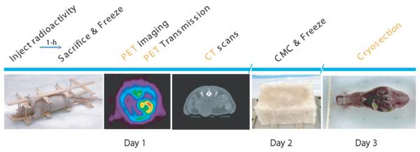
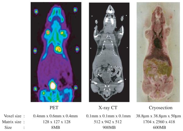
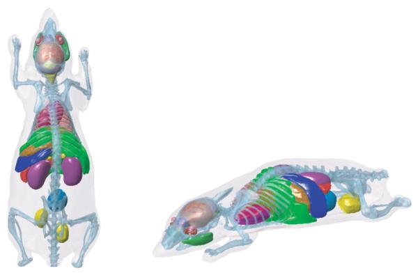
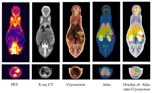
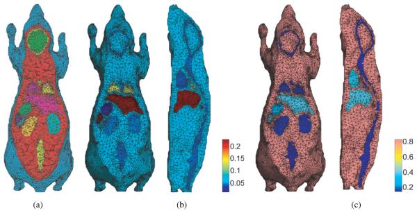
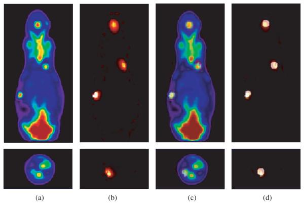
We have constructed a three-dimensional (3D) whole body mouse atlas from coregistered x-ray CT and cryosection data of a normal nude male mouse. High quality PET, x-ray CT and cryosection images were acquired post mortem from a single mouse placed in a stereotactic frame with fiducial markers visible in all three modalities. The image data were coregistered to a common coordinate system using the fiducials and resampled to an isotropic 0.1 mm voxel size. Using interactive editing tools we segmented and labelled whole brain, cerebrum, cerebellum, olfactory bulbs, striatum, medulla, masseter muscles, eyes, lachrymal glands, heart, lungs, liver, stomach, spleen, pancreas, adrenal glands, kidneys, testes, bladder, skeleton and skin surface. The final atlas consists of the 3D volume, in which the voxels are labelled to define the anatomical structures listed above, with coregistered PET, x-ray CT and cryosection images. To illustrate use of the atlas we include simulations of 3D bioluminescence and PET image reconstruction. Optical scatter and absorption values are assigned to each organ to simulate realistic photon transport within the animal for bioluminescence imaging. Similarly, 511 keV photon attenuation values are assigned to each structure in the atlas to simulate realistic photon attenuation in PET. The Digimouse atlas and data are available at http://neuroimage.usc.edu/Digimouse.html.
Figures
Similar articles
-
Estimation of mouse organ locations through registration of a statistical mouse atlas with micro-CT images.IEEE Trans Med Imaging. 2012 Jan;31(1):88-102. doi: 10.1109/TMI.2011.2165294. Epub 2011 Aug 18. IEEE Trans Med Imaging. 2012. PMID: 21859613 Free PMC article.
-
Automated analysis of small animal PET studies through deformable registration to an atlas.Eur J Nucl Med Mol Imaging. 2012 Nov;39(11):1807-20. doi: 10.1007/s00259-012-2188-7. Epub 2012 Jul 21. Eur J Nucl Med Mol Imaging. 2012. PMID: 22820650 Free PMC article.
-
A method of 2D/3D registration of a statistical mouse atlas with a planar X-ray projection and an optical photo.Med Image Anal. 2013 May;17(4):401-16. doi: 10.1016/j.media.2013.02.009. Epub 2013 Mar 5. Med Image Anal. 2013. PMID: 23542374 Free PMC article.
-
X-ray-based attenuation correction for positron emission tomography/computed tomography scanners.Semin Nucl Med. 2003 Jul;33(3):166-79. doi: 10.1053/snuc.2003.127307. Semin Nucl Med. 2003. PMID: 12931319 Review.
-
Positron emission tomography/computed tomography.Semin Nucl Med. 2008 May;38(3):152-66. doi: 10.1053/j.semnuclmed.2008.01.003. Semin Nucl Med. 2008. PMID: 18396176 Review.
Cited by
-
Automated in vivo Assessment of Vascular Response to Radiation using a Hybrid Theranostic X-ray Irradiator/Fluorescence Molecular Imaging System.IEEE Access. 2020;8:93663-93670. doi: 10.1109/access.2020.2994943. Epub 2020 May 15. IEEE Access. 2020. PMID: 32542176 Free PMC article.
-
Vascular supply of the metacarpophalangeal joint.Front Med (Lausanne). 2022 Oct 20;9:1015895. doi: 10.3389/fmed.2022.1015895. eCollection 2022. Front Med (Lausanne). 2022. PMID: 36341235 Free PMC article.
-
Applicability, usability, and limitations of murine embryonic imaging with optical coherence tomography and optical projection tomography.Biomed Opt Express. 2016 May 19;7(6):2295-310. doi: 10.1364/BOE.7.002295. eCollection 2016 Jun 1. Biomed Opt Express. 2016. PMID: 27375945 Free PMC article.
-
Neuronal activity under transcranial radio-frequency stimulation in metal-free rodent brains in-vivo.Commun Eng. 2022;1:15. doi: 10.1038/s44172-022-00014-7. Epub 2022 Jul 1. Commun Eng. 2022. PMID: 38125336 Free PMC article.
-
Recent Technical Advances in Accelerating the Clinical Translation of Small Animal Brain Imaging: Hybrid Imaging, Deep Learning, and Transcriptomics.Front Med (Lausanne). 2022 Mar 24;9:771982. doi: 10.3389/fmed.2022.771982. eCollection 2022. Front Med (Lausanne). 2022. PMID: 35402436 Free PMC article. Review.
References
-
- Broadwell RD, Bleier R. A cytoarchitectonic atlas of the mouse hypothalamus. J. Comp. Neurol. 1976;167:315–39. - PubMed
-
- Burger A, Baldock R, Yang Y. The Edinburgh mouse atlas and gene-expression database: a spatio-temporal database for biological research; Proc. 14th Int. Conf. on Scientific and Statistical Database Management (SSDBM02); 2002. http://genex.hgu.mrc.ac.uk/
-
- Chaudhari AJ, Darvas F, Bading JR, Moats RA, Conti PS, Smith DJ, Cherry SR, Leahy RM. Hyperspectral and multispectral bioluminescence optical tomography for small animal imaging. Phys. Med. Biol. 2005;50:5421–41. - PubMed
Publication types
MeSH terms
Grants and funding
LinkOut - more resources
Full Text Sources
Other Literature Sources