. 2007 Jan;63(1):283-93.
doi: 10.1111/j.1365-2958.2006.05512.x.

Acid pH activation of the PmrA/PmrB two-component regulatory system of Salmonella enterica

Affiliations

Acid pH activation of the PmrA/PmrB two-component regulatory system of Salmonella enterica

J Christian Perez et al. Mol Microbiol. 2007 Jan.

Abstract

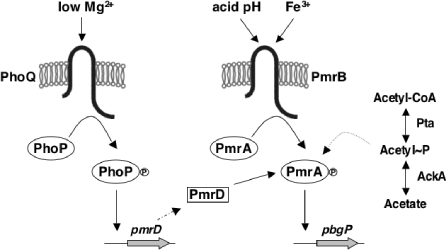
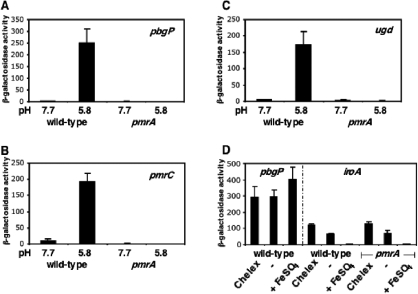
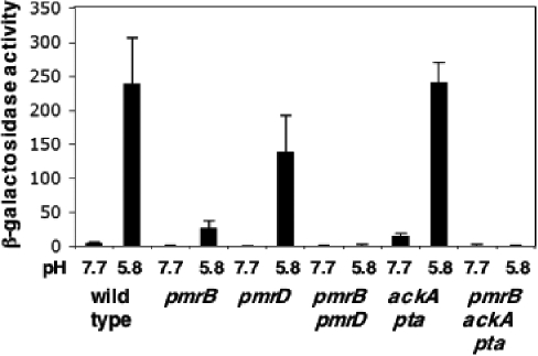
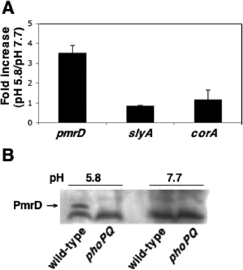
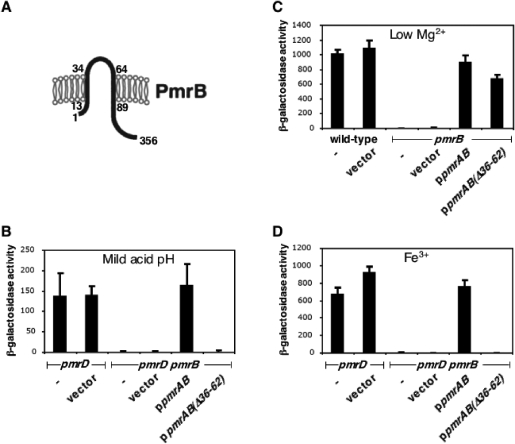
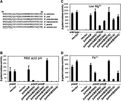
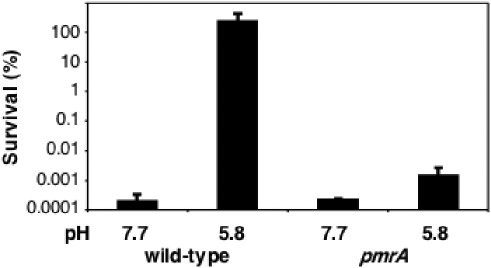
Acid pH often triggers changes in gene expression. However, little is known about the identity of the gene products that sense fluctuations in extracytoplasmic pH. The Gram-negative pathogen Salmonella enterica serovar Typhimurium experiences a number of acidic environments both inside and outside animal hosts. Growth in mild acid (pH 5.8) promotes transcription of genes activated by the response regulator PmrA, but the signalling pathway(s) that mediates this response has thus far remained unexplored. Here we report that this activation requires both PmrA's cognate sensor kinase PmrB, which had been previously shown to respond to Fe(3+) and Al(3+), and PmrA's post-translational activator PmrD. Substitution of a conserved histidine or of either one of four conserved glutamic acid residues in the periplasmic domain of PmrB severely decreased or abolished the mild acid-promoted transcription of PmrA-activated genes. The PmrA/PmrB system controls lipopolysaccharide modifications mediating resistance to the antibiotic polymyxin B. Wild-type Salmonella grown at pH 5.8 were > 100 000-fold more resistant to polymyxin B than organisms grown at pH 7.7. Our results suggest that protonation of the PmrB periplasmic histidine and/or of the glutamic acid residues activate the PmrA protein, and that mild acid promotes cellular changes resulting in polymyxin B resistance.


Figures

Fig. 1
Fig. 1
Model depicting the pathways leading to activation of the PmrA protein. Fe3+ and acid pH are sensed by the PmrB protein, which promotes phosphorylation of PmrA, resulting in transcription of PmrA-activated genes. The PhoQ protein senses extracellular Mg2+. In low Mg2+, PhoQ promotes phosphorylation of PhoP and transcription of pmrD. The PmrD protein binds to the phosphorylated form of PmrA protecting it from dephosphorylation by PmrB. Acetyl phosphate, which is synthesized by the enzymes phosphotransacetylase (Pta) and acetate kinase (AckA), activates PmrA in a strain deleted for the pmrB gene.
Fig. 2
Fig. 2
Mild acid pH promotes transcription of PmrA-regulated genes. A–C. β-Galactosidase activity (Miller units) expressed by strains harbouring chromosomal lac transcriptional fusions to the PmrA-activated pbgP (EG9241, EG9681) (A), pmrC (EG9279, EG9687) (B) and ugd (EG9524, EG9674) (C) genes. Strain numbers are indicated in parenthesis, with the first one corresponding to the pmrA+ and the second to the pmrA background. Expression was investigated in wild-type and pmrA backgrounds following growth in N-minimal medium pH 7.7 or 5.8 as described under Experimental procedures. Shown are the mean values and standard deviations of three independent experiments performed in duplicate. D. β-Galactosidase activity (Miller units) expressed by strains harbouring chromosomal lac transcriptional fusions to the PmrA-activated pbgP (EG9241) and iron-repressed iroA (EG12735, EG12737) genes. Cells were grown in Chelex-treated or untreated N-minimal medium pH 5.8. FeSO4 (100 μM) was added to the Chelex-treated medium where indicated. Shown are the mean values and standard deviations of three independent experiments performed in duplicate.
Fig. 3
Fig. 3
The PmrA-cognate sensor PmrB is required to activate the PmrA-regulated gene pbgP in response to mild acid pH. β-Galactosidase activity (Miller units) expressed by strains harbouring a chromosomal lac transcriptional fusion to the pbgP gene. Expression was investigated in wild-type (EG9241), pmrB (EG16704) and pmrD (EG11775) mutant, pmrB pmrD (EG12060) and ackA pta (EG16450) double mutant and the pmrB ackA pta triple mutant (EG16706) backgrounds. Cells were grown in N-minimal medium pH 7.7 or 5.8 as described under Experimental procedures. Shown are the mean values and standard deviations of three independent experiments performed in duplicate.
Fig. 4
Fig. 4
Expression of the pmrD gene is promoted under mild acid pH. A. RNA levels of transcripts corresponding to the PhoP-activated pmrD and slyA genes and to the PhoP-independent corA gene as determined by quantitative real-time PCR. Shown are the mean values and standard deviations of three independent experiments. B. Western blot analysis of crude bacterial extracts prepared from wild-type (14028s) or phoPQ (EG15598) cells grown in N-minimal medium at pH 5.8 or 7.7 as described under Experimental procedures. The upper band corresponds to PmrD. The lower band is a non-specific cross-reactive product that indicates equal protein loading across the lanes.
Fig. 5
Fig. 5
The periplasmic domain of the PmrB protein is required for responding to mild acid pH. A. Predicted topology of the sensor kinase PmrB in the inner membrane. Numbers indicate amino acid positions. B–D. β-Galactosidase activity (Miller units) expressed by wild type (EG9241), pmrB (EG10065) and pmrD pmrB (EG12060) mutant strains harbouring a lac transcriptional fusion to pbgP and either the plasmid vector pUHE21lacIq, plasmid ppmrAB expressing the wild-type pmrAB genes or plasmid ppmrAB36-62) expressing the wild-type PmrA protein and a PmrB protein deleted for 26 of its 31 periplasmic domain residues. Cells were grown in N-minimal medium containing 10 mM MgCl2, pH 5.8 (B), 10 μM MgCl2, pH 7.7 (C) or 10 μM MgCl2, 100 μM FeSO4, pH 7.7 (D). The β-galactosidase activity in all strains grown under non-inducing conditions, i.e. N-minimal medium containing 10 mM MgCl2, pH 7.7, was undetectable. Shown are the mean values and standard deviations of three independent experiments performed in duplicate.
Fig. 6
Fig. 6
Conserved histidine and glutamic acid residues in the periplasmic domain of the PmrB protein are required for PmrA-mediated transcription in response to mild acid pH. A. Alignment of the amino acid sequences corresponding to the putative periplasmic domains of the PmrB proteins from Salmonella enterica, Escherichia coli, Klebsiella pneumoniae, Serratia marcescens, Yersinia pestis and Erwinia carotovora. Asterisks (*) denote residues conserved in all six species. B–D. β-Galactosidase activity (Miller units) expressed by wild-type (EG9241), pmrB (EG10065) and pmrD pmrB (EG12060) mutant strains harbouring a lac transcriptional fusion to pbgP and plasmid vector pUHE21lacIq, plasmid ppmrAB expressing the wild-type pmrAB genes, or plasmids in which the nucleotide sequence corresponding to periplasmic histidines and glutamates were mutated to alanine [ppmrAB(H35A), ppmrAB(E36A), ppmrAB(E39A), ppmrAB(H57A), ppmrAB(E61A), ppmrAB(E64A)]. Cells were grown in N-minimal medium containing 10 mM MgCl2, pH 5.8 (B), 10 μM MgCl2, pH 7.7 (C) or 10 μM MgCl2, 100 μM FeSO4, pH 7.7 (D). The β-galactosidase activity in all strains grown under non-inducing conditions, i.e. N-minimal medium containing 10 mM MgCl2, pH 7.7, was undetectable. Shown are the mean values and standard deviations of three independent experiments performed in duplicate.
Fig. 7
Fig. 7
Mild acid pH induces resistance to the antimicrobial peptide polymyxin B. Per cent survival of wild-type (14028s) and pmrA (EG7139) strains after incubation with polymyxin B (1.5 μg ml−1). Cells were grown in N-minimal medium, pH 7.7 or 5.8, containing 10 mM MgCl2, before incubation with polymyxin B. Shown are the mean values and standard deviations of three independent experiments performed in duplicate.

Similar articles

Cited by

References

    1. Aranda CMA, Swanson JA, Loomis WP, Miller SI. Salmonella typhimurium activates virulence gene transcription within acidified macrophage phagosomes. Proc Natl Acad Sci USA. 1992;89:10079–10083. - PMC - PubMed
    1. Bearson BL, Wilson L, Foster JW. A low pH-inducible, PhoPQ-dependent acid tolerance response protects Salmonella typhimurium against inorganic acid stress. J Bacteriol. 1998;180:2409–2417. - PMC - PubMed
    1. Breazeale SD, Ribeiro AA, Raetz CRH. Origin of lipid A species modified with 4-amino-4-deoxy-l-arabinose in polymyxin-resistant mutants of Escherichia coli: an aminotransferase (ArnB) that generates UDP-4-amino-4-deoxy-l-arabinose. J Biol Chem. 2003;278:24731–24739. - PubMed
    1. Brumell JH, Grinstein S. Salmonella redirects phagosomal maturation. Curr Opin Microbiol. 2004;7:78–84. - PubMed
    1. Chamnongpol S, Dodson W, Cromie MJ, Harris ZL, Groisman EA. Fe(III)-mediated cellular toxicity. Mol Microbiol. 2002;45:711–719. - PubMed

Publication types

MeSH terms