. 2007 Jan;210(1):66-77.
doi: 10.1111/j.1469-7580.2006.00674.x.

Insights into the gyrification of developing ferret brain by magnetic resonance imaging

Affiliations

Insights into the gyrification of developing ferret brain by magnetic resonance imaging

Jason Neal et al. J Anat. 2007 Jan.

Abstract

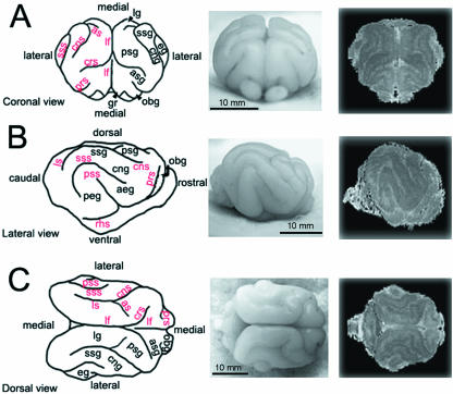
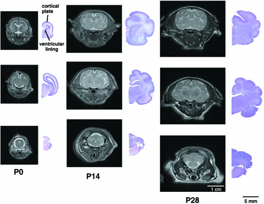
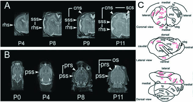
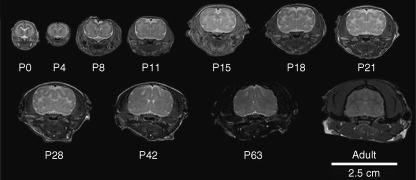
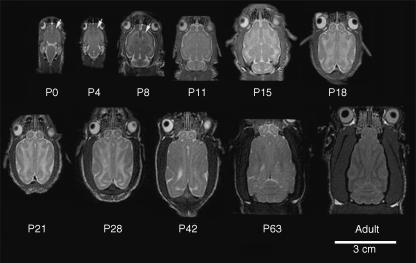
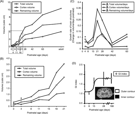
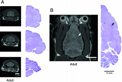
The developmental mechanisms underlying the formation of human cortical convolutions (gyri and sulci) remain largely unknown. Genetic causes of lissencephaly (literally 'smooth brain') would imply that disorders in neuronal migration cause the loss of cortical convolutions. However, prior studies have suggested that loss of sulci and gyri can also arise from impaired proliferation, disrupted lamination and loss of intracortical connections. To gain further insight into the mechanisms underlying the formation of cortical convolutions, we examined the progressive brain development of the gyrencephalic ferret. In this study, we used magnetic resonance imaging to follow the temporal and spatial pattern of neuronal migration, proliferation and differentiation in relation to the onset and development of cortical convolutions. In this manner, we demonstrate that the onset of gyrification begins largely after completion of neuronal proliferation and migration. Gyrification occurs in a lateral to medial gradient, during the period of most rapid cerebral cortical growth. Cortical folding is also largely complete prior to myelination of the underlying cortical axons. These observations are consistent with gyrification arising secondary to cortical processes involving neuronal differentiation.


Figures

Fig. 1
Fig. 1
Gross anatomical profiles of the ferret brain at 4 weeks of age. Camera lucida renderings (left), photomicrographs (centre), and 3D reconstructions of the 4-week-old ferret brain are displayed in coronal (A), lateral (B) and dorsal (C) views. The prominent, rounded elevations found on the surface of the brain tissue (gyri) with their corresponding acronyms are denoted in black. The furrows along the surface of the brain (sulci) with their corresponding acronyms are denoted in red. The actual sulcal and gyral labels for these abbreviations are listed in Table 2.
Fig. 2
Fig. 2
Sulcal and gyral folds in the ferret brain form postnatally. Rostral to caudal coronal MR images (left) with corresponding cresyl violet sections (right) are shown from various aged ferret brains (postnatal day 0, P14, P28).
Fig. 3
Fig. 3
Ferret sulcal and gyral formation progress along a lateral to medial gradient. (A) Coronal MR images of early developmental time points show initial development of the more lateral rhinal sulcus, followed by the appearance of the more medial suprasylvian sulci and finally, the most medially positioned coronal and splenocruciate sulci. (B) Axial MR images of early developmental time points similarly display a lateral to medial progression of sulcal development, beginning with the pseudosylvian sulcus and later followed by development of the more medially positioned presylvian sulcus and the most medial olfactory sulcus. (C) Camera lucida drawings (coronal, lateral and dorsal views) of the 4-week brain surface display the overall lateral to medial progression of sulcal and gyral development (arrows). The SCS (asterisk) is obscured and lies ventral and lateral to the longitudinal fasiculus (lf).
Fig. 4
Fig. 4
Neuronal proliferation and migration in postnatal ferret by MR imaging. Coronal MR images of the ferret brain were obtained at various ages [P0 to adulthood (6 months)]. At P0, the T2-weighted dark signal located along lateral ventricular lining (arrow) suggests the presence of a high density of progenitor cells. At P4 and P8, this dark contrast progressively diminishes along the ventricles but increases at the outer cortical surface (arrowhead). This corresponds to the gradual completion of neuronal proliferation at the ventricular zone and the progression of postmitotic neuronal migration from the ventricular zone to the cortical plate during the first postnatal week.
Fig. 5
Fig. 5
Oligodendrocyte myelination in postnatal ferret by MR imaging. Serial T2-weighted axial MR images of the ferret brain were obtained at various ages (birth until adulthood). The same T2 dark labelling (arrows) seen along the ventricular lining and later in the cortical plate in the coronal sections is appreciated on the axial views during the first postnatal week, probably corresponding to the migration of immature neuroblasts from the VZ to the cortical plate. From P0 to P28, rapid brain expansion is accompanied by an increase in T2 brightening beneath the cortical plate, consistent with progressive elaboration of axonal projections. Thereafter, axial images display gradual tapering of the rostral forebrain and an increase in T2 darkening beneath the cortical plate, consistent with an increase in axonal fibre tract myelination, and a decrease in the overall number of axonal tracts in the brain.
Fig. 6
Fig. 6
Gyrification occurs during the period of greatest increase in cerebral cortical volume. (A) The volume of ferret brain structures (cortex, subcortical structures, entire brain) shows the greatest increase between the 2nd and 4th weeks of postnatal development. (B) Magnified view of the inset in (A) shows that the cortical volume actually surpasses the volume of the subcortical structures by the fourth postnatal day (asterisk). This time frame corresponds to the progressive migration of neurons from the ventricular zone to the cortical plate. (C) The cortical growth rate exceeds both the growth rate of the total brain and its underlying structures (remaining volume) in the first postnatal week (asterisk), consistent with expansion of the cortical plate. Thereafter, the cortex undergoes a sustained period of growth between the second and fourth weeks of growth. (D) Gyrification index indicates that gyrification advances most rapidly during the third postnatal week, consistent with the period of most rapid cortical growth. Subsequently, the gyral formation plateaus until adulthood. Image displays a manually traced P28 coronal section used to calculate GI.
Fig. 7
Fig. 7
Increase in brain size from axonal myelination does not contribute to sulcal and gyral formation. (A) Serial T2-weighted coronal MR images and corresponding cresyl violet brain sections from adult ferret brain at 6 months of age. The now densely myelinated fibre tracts are T2 dark and appear denser than the overlying cortical structures and deeper basal structures. The cerebral cortex has become less dense (T2 dark) with increasing neuronal maturation. (B) A single axial MR image at the level of the interventricular foramen. The densely myelinated fibres (black arrowhead) on cresyl violet staining corresponds to the T2 dark signal (white arrowhead) on the MR image. Although the overall size of the ferret brain has increased between 4 weeks and 6 months of age as a result of axonal myelination, there is no change in the sulcal and gyral patterns.

References

    1. Benveniste H, Blackband S. MR microscopy and high resolution small animal MRI: applications in neuroscience research. Prog Neurobiol. 2002;67:393–420. - PubMed
    1. Clark GD. Cerebral gyral dysplasias: molecular genetics and cell biology. Curr Opin Neurol. 2001;14:157–162. - PubMed
    1. Fox JG. Biology and Diseases of the Ferret. Baltimore, MD: Lippincott, Williams & Wilkins; 1998.
    1. Francis F, Meyer G, Fallet-Bianco C, et al. Human disorders of cortical development: from past to present. Eur J Neurosci. 2006;23:877–893. - PubMed
    1. Hilgetag CC, Barbas H. Developmental mechanics of the primate cerebral cortex. Anat Embryol (Berl) 2005;210:411–417. - PubMed

Publication types

MeSH terms