. 2007 May;19(5):1056-70.
doi: 10.1016/j.cellsig.2006.12.001. Epub 2006 Dec 12.

Palmitoylation of the TPbeta isoform of the human thromboxane A2 receptor. Modulation of G protein: effector coupling and modes of receptor internalization

Affiliations

Palmitoylation of the TPbeta isoform of the human thromboxane A2 receptor. Modulation of G protein: effector coupling and modes of receptor internalization

Helen M Reid et al. Cell Signal. 2007 May.

Erratum in

Abstract

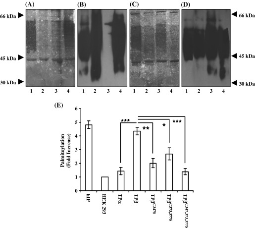
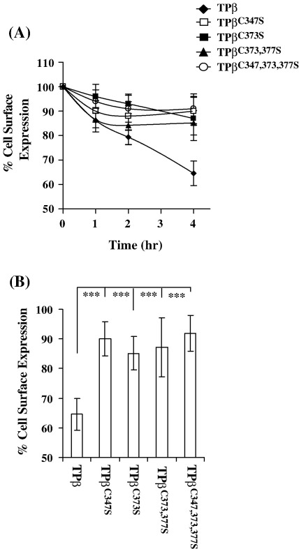
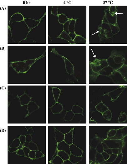
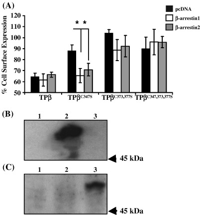
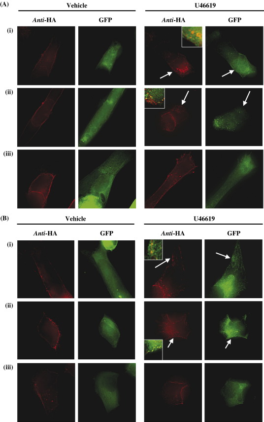
Palmitoylation is a prevalent feature amongst G protein-coupled receptors. In this study we sought to establish whether the TPalpha and TPbeta isoforms of the human prostanoid thromboxane (TX) A2 receptor (TP) are palmitoylated and to assess the functional consequences thereof. Consistent with the presence of three cysteines within its unique carboxyl-terminal domain, metabolic labelling and site-directed mutagenesis confirmed that TPbeta is palmitoylated at Cys347 and, to a lesser extent, at Cys373,377 whereas TPalpha is not palmitoylated. Impairment of palmitoylation did not affect TPbeta expression or its ligand affinity. Conversely, agonist-induced [Ca2+]i mobilization by TPbetaC347S and the non-palmitoylated TPbetaC347,373,377S, but not by TPbetaC373S or TPbetaC373,377S, was significantly reduced relative to the wild type TPbeta suggesting that palmitoylation at Cys347 is specifically required for efficient Gq/phospholipase Cbeta effector coupling. Furthermore, palmitoylation at Cys373,377 is critical for TPbeta internalization with TPbetaC373S, TPbetaC373,377S and TPbetaC347,373,377S failing to undergo either agonist-induced or temperature-dependent tonic internalization. On the other hand, whilst TPbetaC347S underwent reduced agonist-induced internalization, it underwent tonic internalization to a similar extent as TPbeta. The deficiency in agonist-induced internalization by TPbetaC347S, but not by TPbetaC373,377 nor TPbeta(C347,373,377S), was overcome by over-expression of either beta-arrestin1 or beta-arrestin2. Taken together, data herein suggest that whilst palmitoylation of TPbeta at Cys373,377 is critical for both agonist- and tonic-induced internalization, palmitoylation at Cys347 has a role in determining which pathway is followed, be it by the beta-arrestin-dependent agonist-induced pathway or by the beta-arrestin-independent tonic internalization pathway.


Figures

Fig. 1
Fig. 1
Analysis of palmitoylation in HEK.TPα and HEK.TPβ cells. Panels A and B, HEK.TPβ (lane 1), HEK.TPα (lane 2), HEK 293 (lane 3, negative control) and HEK.hIP (lane 4, positive control) cells were metabolically labelled for 2 h at 37 °C (Panel A). Panels C and D, HEK.TPβ (lane 1), HEK.TPβC347S (lane 2), HEK.TPβC373,377S cells (lane 3) and HEK.TPβC347,373,377S (lane 4) cells were metabolically labelled for 2 h at 37 °C (Panel C). Thereafter, the HA-tagged receptors were immunoprecipitated with anti-HA 101R and resolved by SDS-PAGE followed by electroblotting onto PVDF membrane. The blots (A and C) were soaked in Amplify prior to fluorography for 60–90 days at − 70 °C. Following fluorographic exposure, PDVF membranes in A and C were screened by immunoblot analysis using anti-HA 3F10 peroxidase-conjugated antibody followed by chemiluminescent detection to obtain Panels B and D, respectively. The positions of the molecular weight markers (kDa) are indicated to the left and right of Panels A and D, respectively. Panel E, the level of palmitoylation in HEK.hIP, HEK.TPα, HEK.TPβ, HEKHA.TPβC347S, HEK.TPβC373,377S and HEK.TPβC347,373,377S cells relative to basal levels, in HEK 293 cells, was determined by Phosphorimage analysis. Data is presented as mean fold increase in palmitoylation over basal levels ± S.E.M. and are expressed in arbitrary units. The data are representative of three independent experiments. The asterisks indicate that palmitoylation levels of TPα, TPβC347S, TPβC373,377S and TPβC347,373,377S cells were significantly lower than that of TPβ where , p < 0.05; ⁎⁎, p < 0.01; ⁎⁎⁎p < 0.005.
Fig. 2
Fig. 2
Analysis of U46619-mediated [Ca2+]i mobilization. HEK.TPβ (Panel A), HEK.TPβC347S (Panel B), HEK.TPβC373S (Panel C), HEK.TPβC373,377S (Panel D) and HEK.TPβC347,373,377S (Panel E) cells, transiently co-transfected with pCMV5:Gαq and preloaded with FURA2/AM, were stimulated with 1 μM U46619 where the ligand was added at times indicated by the arrows. The results are representative profiles from 4 independent experiments and are plotted as changes in intracellular Ca2+ mobilized (Δ[Ca2+]i, nM) as a function of time(s) following ligand stimulation. Data presented in Panel F is plotted as mean changes in Δ[Ca2+]i mobilization (nM ± S.E.M.) for each cell line. The asterisk in Panel F indicates that U46619-mediated [Ca2+]i mobilization by TPβC347S and TPβC347,373,377S was significantly lower than that of TPβ where , p < 0.05.
Fig. 3
Fig. 3
Effect of palmitoylation on agonist-induced internalization of TPβ. Panel A, HEK.TPβ, HEK.TPβC347S, HEKHA.TPβC373S, HEK.TPβC373,377S and HEK.TPβC347,373,377S cells were incubated with 1 μM U46619 at 37 °C for 0–4 h prior to fixation and immunolabelling with anti-HA 101R. Results are expressed as mean cell surface expression at each time point as a percentage of that at 0 h (% cell surface expression ± S.E.M., n = 3) as a function time (h). Panel B, mean cell surface expression following stimulation of cells with 1 μM U46619, 37 °C for 4 h expressed as a percentage of that at 0 h (% cell surface expression ± S.E.M., n = 3). The asterisks indicate that at 4 h, U46619-mediated loss of cell surface expression of TPβC347S, TPβC373S, TPβC373,377S and TPβC347,373,377S was significantly reduced relative to that of TPβ where ⁎⁎⁎, p < 0.0001.
Fig. 4
Fig. 4
Confocal microscopy of agonist-induced internalization. HEK.TPβ (Panel A), HEK.TPβC347S (Panel B), HEK.TPβC373,377S (Panel C) and HEK.TPβC347,373,377S (Panel D) cells were with pre-labelled with anti-HA 101R, at 4 °C for 1 h, prior to stimulation with 1 μM U46619 at 37 °C for the times indicated. Thereafter, following fixation, surface expression of the HA-tagged receptors was detected, under both non-permeabilizing and permeabilizing conditions, by immunolabelling anti-mouse FITC-conjugated antibody. Images were captured using a Carl Zeiss Lazer Scanning System LSM510 and Zeiss LSM Imaging software. The arrows show agonist-induced TPβ internalization as indicated by loss of cell surface HA-tagged TPβ detected in non-permeabilized cells and increased detection of intracellular TPβ in permeabilized cells. The colour version of this figure is available online at www.sciencedirect.com.
Fig. 5
Fig. 5
Effect of palmitoylation on tonic internalization. HEK.TPβ, HEK.TPβC347S, HEK. TPβC373S, HEK.TPβC373,377S and HEK.TPβC347,373,377S cells were incubated in the presence of anti-HA 101R for 1 h at 4 °C. Thereafter, following washing, cells were incubated at 4 °C or 37 °C for 2 h. Following fixation, cell surface expression of the HA-tagged receptors was detected using anti-mouse HRP conjugate antibody followed by colorimetric detection. Results are expressed as cell surface expression at 2 h as percentage of that at 0 h at the respective temperatures (% cell surface expression ± S.E.M., n = 3). The asterisks indicates that at 37 °C, tonic internalization of TPβC373S, TPβC373,377S and TPβC347,373,377S was significantly reduced compared to that of TPβ where , p < 0.0001 and ⁎⁎, p < 0.0005.
Fig. 6
Fig. 6
Confocal microscopy of tonic internalization. HEK.TPβ (Panel A), HEK.TPβC347S (Panel B), HEK.TPβC373,377S (Panel C) and HEK.TPβC347,373,377S (Panel D) cells were pre-labelled with anti-HA 101R at 4 °C for 1 h. Thereafter, following washing, cells were incubated for 2 h at 4 °C or 37 °C, followed by fixation in paraformaldehyde. Cell surface expression of the HA-tagged receptors was detected using anti-mouse FITC-conjugated antibody, under permeabilizing conditions. Images were captured using a Carl Zeiss Lazer Scanning System LSM510 and Zeiss LSM Imaging software. The arrows show temperature-induced tonic internalization of TPβ and TPβC347S. The colour version of this figure is available online at www.sciencedirect.com.
Fig. 7
Fig. 7
Effect of β-arrestin on agonist-induced internalization. Panel A, HEK.TPβ, HEK.TPβC347S, HEK.TPβC373S, HEK.TPβC373,377S and HEK.TPβC347,373,377S cells, transiently co-transfected with pRK5.β-arrestin1, pcDNA1.β-arrestin2 or, as a control, with the empty vector pcDNA were incubated with U46619 (1 μM) at 37 °C for 4 h, prior to fixation and immunolabelling with anti-HA 101R. Following fixation, cell surface expression of the HA-tagged receptors was detected using anti-mouse HRP-conjugated antibody followed by colorimetric detection. Results are expressed as mean cell surface expression at 4 h as a percentage of that at 0 h (% cell surface expression ± S.E.M., n = 3). The asterisk (, p < 0.05) indicates that over-expression of β-arrestin1 and β-arrestin2 significantly reduced the level of cell surface expression of TPβC347S in the presence of U46619. Panels B and C, HEK.TPβ cells, transiently co-transfected with pRK5.β-arrestin1 (lane 2), pcDNA1.β-arrestin2 (lane 3) or, as a control, with the empty vector pcDNA (lane 1) were analysed by SDS-PAGE (50 μg whole cell protein analysed/lane) followed by Western blot analysis using anti-β-arrestin1 (K-16: Panel B) and anti-β-arrestin2 (N-16: Panel C). Data presented are representative immunoblots from three independent experiments. The relative position of the 45 kDa molecular size marker is indicated to the right of Panels B and C.
Fig. 8
Fig. 8
Effect of palmitoylation on TPβ and β-arrestin co-localization. Panels A and B, HEK 293 cells transiently co-transfected with (i) pHM6.TPβ, (ii) TPβC347S and (iii) TPβC347,373,377S along with pEGFPN3:β-arrestin1 (Panel A) or pEGFPN1:β-arrestin2 (Panel B) were immunolabelled with anti-HA 101R for 1 h at 4 °C. Thereafter, cells incubated with vehicle or U46619 (1 μM) for 30 min at 37 °C. Following fixation and permeabilization, cell surface HA-tagged TPs were labelled with the AlexaFluor 594 anti-mouse IgG. Images were captured using the Zeiss Fluorescence microscope with AxioVision software (version 4.4) and processed by AxioVision Deconvolution analysis. The arrows show agonist-induced β-arrestin1 and β-arrestin2 translocation and TPβ internalization while the insets to the panels show the co-localization of the β-arrestins with the TPβ receptors. The colour version of this figure is available online at www.sciencedirect.com.
Fig. 9
Fig. 9
Model of palmitoylation of TPβ. Palmitoylation of TPβ occurs at 2 distinct sites through the attachment of palmitates at Cys347 and Cys373/377. Following palmitoylation, it is proposed that membrane insertion of the palmitate moieties at Cys347 and Cys373/377 leads to the formation of a double loop structure within the C-tail domain of TPβ referred to as Loop A and Loop B, respectively. A third conformation of the C-tail of TPβ occurs when Cys373,377, but not Cys347, is palmitoylated resulting in the formation of Loop C, such as is found in TPβC347S. TPβC373,377S undergoes palmitoylation at Cys347 resulting in the formation of Loop A only while TPβC347,373,377S does not contain Loops A, B or C. It was found that Loop A is required for efficient Gαq/PLCβ coupling as evidenced by the reduced agonist-induced [Ca2+]i mobilization by TPβC347S and TPβC347,373,377S. It is proposed that Loop B is required for agonist-induced GRK phosphorylation at Ser357, followed by β-arrestin recruitment and TPβ internalization, as evidenced by the impaired U46619-induced β-arrestin1/2 recruitment and internalization by TPβC347S, TPβC373,377S and TPβC347,373,377S. Like the wild type TPβ, TPβC347S (which contains Loop C only) retains the ability to undergo tonic internalization while TPβC373,377S (which contains Loop A only) or TPβC347,373,377S fails to undergo tonic or agonist-induced internalization. From these latter data, it is proposed that palmitoylation at Cys373 and/or Cys377, resulting in the formation of Loop C only (in TPβC347S) or Loop A plus Loop B (in wild type TPβ) is necessary to present a tonic internalization motif, such as the proposed Y339X3Φ343, in the correct orientation for interaction with the internalization machinery.

Similar articles

Cited by

References

    1. Milligan G., Parenti M., Magee A.I. Trends Biochem. Sci. 1995;20(5):181. - PubMed
    1. Dunphy J.T., Linder M.E. Biochim. Biophys. Acta. 1998;1436(1–2):245. - PubMed
    1. Bijlmakers M.J., Marsh M. Trends Cell Biol. 2003;13(1):32. - PubMed
    1. Qanbar R., Bouvier M. Pharmacol. Ther. 2003;97(1):1. - PubMed
    1. Torrecilla I., Tobin A.B. Curr. Pharm. Des. 2006;12(14):1797. - PubMed

Publication types

MeSH terms

LinkOut - more resources