. 2007 Apr;81(7):3447-54.
doi: 10.1128/JVI.02097-06. Epub 2007 Jan 17.

Formation of a multiple protein complex on the adenovirus packaging sequence by the IVa2 protein

Affiliations

Formation of a multiple protein complex on the adenovirus packaging sequence by the IVa2 protein

Ryan E Tyler et al. J Virol. 2007 Apr.

Abstract

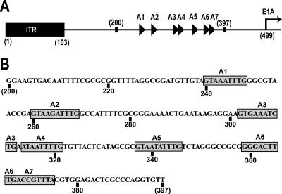
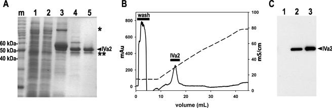
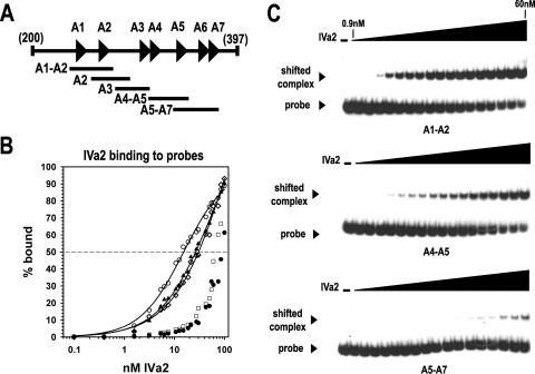
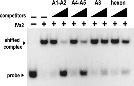
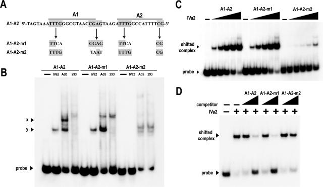
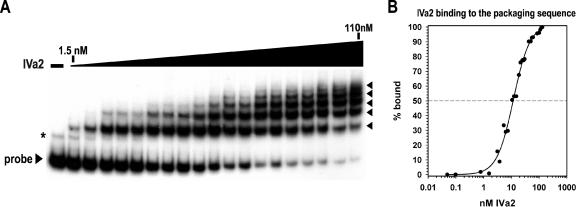
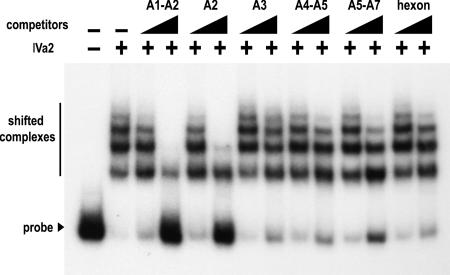
During adenovirus virion assembly, the packaging sequence mediates the encapsidation of the viral genome. This sequence is composed of seven functional units, termed A repeats. Recent evidence suggests that the adenovirus IVa2 protein binds the packaging sequence and is involved in packaging of the genome. Study of the IVa2-packaging sequence interaction has been hindered by difficulty in purifying the protein produced in virus-infected cells or by recombinant techniques. We report the first purification of a recombinant untagged version of the adenovirus IVa2 protein and characterize its binding to the packaging sequence in vitro. Our data indicate that there is more than one IVa2 binding site within the packaging sequence and that IVa2 binding to DNA requires the A-repeat consensus, 5'-TTTG-(N(8))-CG-3'. Furthermore, we present evidence that IVa2 forms a multimeric complex on the packaging sequence. These data support a model in which adenovirus DNA packaging occurs via the formation of a IVa2 multiprotein complex on the packaging sequence.


Figures

FIG. 1.
FIG. 1.
(A) Arrangement of the left end of the Ad5 genome. The A repeats, represented as triangles, are located between nucleotides 200 and 397, upstream of the transcription start site of the E1A promoter, shown as a right-facing arrow. Modified with permission from P. Hearing. (B) Ad5 sequence from nucleotide 200 to 397. The A repeats are highlighted with gray boxes. Specific nucleotide numbers are located below the sequence.
FIG. 2.
FIG. 2.
Expression and purification of recombinant, untagged IVa2 protein. (A) ProtoBlue-stained 9% SDS-PAGE. Intein-tagged IVa2 protein was affinity purified from soluble E. coli extract with a chitin agarose matrix as described in Materials and Methods. Lanes: m, protein standard marker; 1, soluble extract from uninduced E. coli; 2, soluble extract from isopropyl-β-d-thiogalactopyranoside-induced E. coli; 3, boiled intein-tagged IVa2 protein-bound chitin matrix beads; 4, 5 μg of IVa2 protein eluted from the chitin matrix; 5, 5 μg of IVa2 protein eluted from the Mono S PE 4.5/100 column. The purified full-length IVa2 protein is indicated with a labeled arrowhead. *, intein-tagged fusion IVa2 protein; **, IVa2 protein degradation product. (B) Typical chromatogram of the elution profile of the IVa2 protein from a Mono S column. The optical density at 280 nm (mAU) and ionic strength (mS/cm) of the buffer eluted from the column are represented as a solid and a dashed line, respectively. (C) Western blot comparing the IVa2 protein and IVa2 protein from infected cell lysates. Lanes: 1, 15 μg of 293 whole cell lysate; 2, 15 μg of Ad5-infected 293 whole cell lysate; 3, 150 ng of IVa2.
FIG. 3.
FIG. 3.
IVa2 binding to packaging sequence probes. (A) Diagram of the packaging sequence region from nucleotide 200 to 397. The specific overlapping probes are indicated below the complete packaging sequence. (B) Binding of IVa2 to the specific probes. The percentages of the DNA probes bound by IVa2 (% bound) were calculated using PhosphorImager scanning data from the averages of three to four experiments. Symbols: ○, A1-A2 probe; ▴, A2 probe; ⋄, A4-A5 probe; □, A5-A7 probe; •, A3 probe. The fitting of a sigmoidal function to the data is shown as a solid line (see Material and Methods). (C) Representative autoradiograms from EMSAs using the A1-A2, A4-A5, and A5-A7 radiolabeled probes. The dashes above the first lanes indicate that the reaction mixture lacks IVa2. The triangles above the lanes represent increasing concentrations of IVa2 (0.1, 0.4, 1.6, 3.1, 5.3, 6.3, 7.5, 10.6, 12.5, 15, 18.8, 21.3, 27.5, 30, 42.5, 50, and 60 nM). The IVa2-DNA complex and the free probe are indicated with labeled arrowheads.
FIG. 4.
FIG. 4.
Competition assay of IVa2 binding to the A1-A2 probe. EMSAs were performed as described in the legend to Fig. 3 with 40 nM IVa2 and a radiolabeled A1-A2 probe. EMSA binding reaction mixtures were incubated with or without a 20- or 200-fold molar excess of unlabeled DNA encoding the A1-A2 repeats, the A4-A5 repeats, the A3 repeat, or an equal length of DNA from the hexon coding region. The presence or absence of IVa2 in the binding reaction mixture is indicated with a + or −, respectively. The first two lanes lack probes. The triangles above the lanes indicate increasing concentrations of the competitors.
FIG. 5.
FIG. 5.
Interaction of IVa2 with mutant A1-A2 probes. (A) Sequences of the wild-type and mutant probes. The A1-repeat bipartite consensus, 5′-TTTG-(N8)-CG-3′, is indicated by bold lines over the sequence, with the conserved nucleotide motifs in gray boxes. (B) EMSAs comparing binding of A1-A2 wild-type and mutant radiolabeled probes to either 60 nM recombinant IVa2 (IVa2) or 4 μg nuclear extract from adenovirus type 5-infected (Ad5) and mock-infected (293) 293 cells. The dash above the first lane of each set indicates that the reaction mixture contains no IVa2 or extract. The labeled arrowheads indicate the formation of Ad5-specific complexes x and y on the A1-A2 wild-type probe. (C) EMSAs with the A1-A2 wild-type and mutant radiolabeled probes. The dash above the first lane of each set indicates that the reaction mixture contains no IVa2. The triangles above the lanes represent increasing concentrations of IVa2 (3.1, 6.3, 12.5, 25, and 50 nM). (D) Representative autoradiograms from competition assays of IVa2 protein binding to radiolabeled A1-A2. EMSAs were performed as described in the legend to Fig. 4, using the indicated competitors. The IVa2 protein-DNA-shifted complex and free probe are indicated with labeled arrowheads. The presence or absence of IVa2 in the binding reaction is indicated with a + or −, respectively. The first two lanes lack competitor. The triangles above the lanes indicate increasing concentrations of the competitors.
FIG. 6.
FIG. 6.
Multiple complexes form as IVa2 interacts with the packaging sequence. (A) Representative autoradiogram from an EMSA using a radiolabeled probe to nucleotide 200 to 397. The first lane lacks IVa2. The triangle above the lanes represents increasing concentrations of IVa2 (1.5, 3.1, 5.3, 6.3, 7.5, 10.6, 12.5, 15, 18.8, 21.3, 27.5, 30, 42.5, 50, 55, 60, 85, 100, and 110 nM). The IVa2-packaging sequence complexes are indicated with arrowheads. A nonspecific band that was consistently observed in the preparation of the complete packaging sequence probe is indicated with an asterisk. (B) Binding of IVa2 to the packaging sequence probe. The percentage of probe bound by IVa2 (% bound) was calculated as described in the legend to Fig. 3.
FIG. 7.
FIG. 7.
Competition assay of IVa2 binding to the packaging sequence probe. EMSAs were performed as described in the legend to Fig. 4, using either a 100 or 1,000-fold molar excess of the indicated unlabeled A-repeat DNA competitors. The IVa2-packaging sequence probe complexes and the free probe are indicated with a bold line and an arrowhead, respectively. The presence or absence of IVa2 in the binding reaction is indicated with a + or −, respectively. The first two lanes lack competitor. The triangles above the lanes indicate increasing concentrations of the competitors.

References

    1. Anderson, C. W., P. R. Baum, and R. F. Gesteland. 1973. Processing of adenovirus 2-induced proteins. J. Virol. 12:241-252. - PMC - PubMed
    1. Anderson, D., and S. Grimes. 2005. The Φ29 DNA packaging motor, p. 102-116. In C. E. Catalano (ed.), Viral genome packaging machines: genetics, structure, and mechanism. Landes Bioscience/Eurekah.com, Georgetown, TX.
    1. Daniell, E. 1976. Genome structure of incomplete particles of adenovirus. J. Virol. 19:685-708. - PMC - PubMed
    1. Edvardsson, B., E. Everitt, H. Jornvall, L. Prage, and L. Philipson. 1976. Intermediates in adenovirus assembly. J. Virol. 19:533-547. - PMC - PubMed
    1. Feiss, M., and C. E. Catalano. 2005. Bacteriophage lambda terminase and mechanisms of DNA packaging, p. 5-39. In C. E. Catalano (ed.), Viral genome packaging machines: genetics, structure and mechanism. Landes Bioscience/Eurekah.com, Georgetown, TX.

Publication types

LinkOut - more resources