. 2007 Apr;81(7):3377-90.
doi: 10.1128/JVI.02191-06. Epub 2007 Jan 17.

Maintenance of endoplasmic reticulum (ER) homeostasis in herpes simplex virus type 1-infected cells through the association of a viral glycoprotein with PERK, a cellular ER stress sensor

Affiliations

Maintenance of endoplasmic reticulum (ER) homeostasis in herpes simplex virus type 1-infected cells through the association of a viral glycoprotein with PERK, a cellular ER stress sensor

Matthew Mulvey et al. J Virol. 2007 Apr.

Abstract

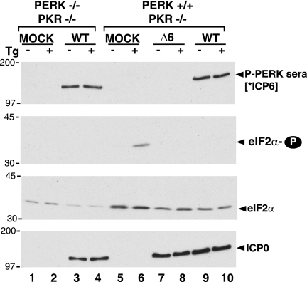
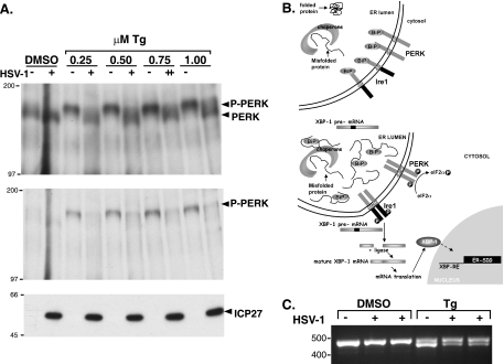
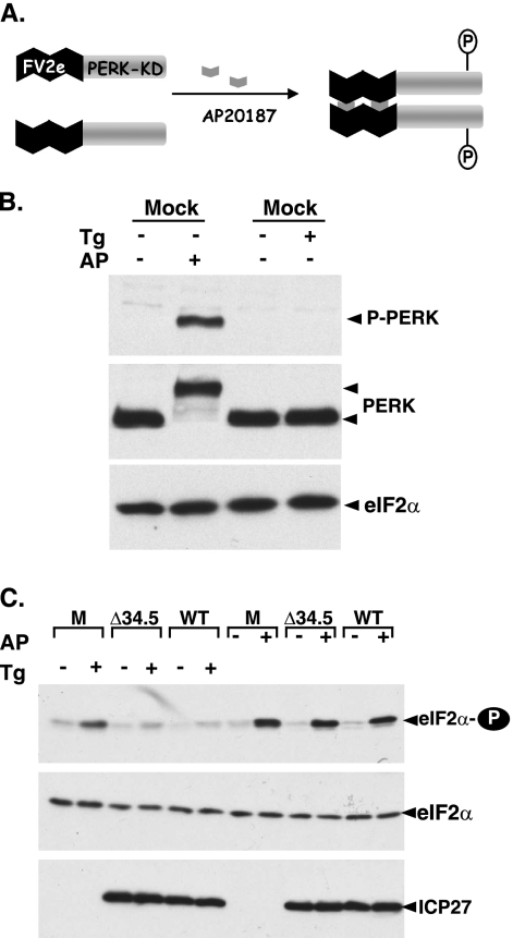
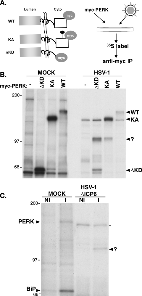
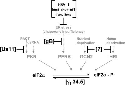
In the efforts of viruses to dominate and control critical cellular pathways, viruses generate considerable intracellular stress within their hosts. In particular, the capacity of resident endoplasmic reticulum (ER) chaperones to properly process the acute increase in client protein load is significantly challenged. Such alterations typically induce the unfolded protein response, one component of which acts through IRE1 to restore ER homeostasis by expanding the folding capabilities, whereas the other arm activates the eIF-2alpha (alpha subunit of eukaryotic initiation factor 2) kinase PERK to transiently arrest production of new polypeptide clientele. Viruses, such as herpes simplex virus type 1 (HSV-1), however, go to great lengths to prevent the inhibition of translation resulting from eIF-2alpha phosphorylation. Here, we establish that PERK, but not IRE1, resists activation by acute ER stress in HSV-1-infected cells. This requires the ER luminal domain of PERK, which associates with the viral glycoprotein gB. Strikingly, gB regulates viral protein accumulation in a PERK-dependent manner. This is the first description of a virus-encoded PERK-specific effector and defines a new strategy by which viruses are able to maintain ER homeostasis.


Figures

FIG. 1.
FIG. 1.
Cross-reaction of phospho-PERK antiserum with the HSV-1 ICP6 polypeptide. Immortalized mouse embryonic fibroblasts (PERK−/− PKR−/− or PERK+/+ PKR−/−) were mock infected (MOCK) or infected with HSV-1 (MOI of 5) (wild-type [WT] or an ICP6-deficient strain [Δ6]). At 9 hpi, cultures were treated with thapsigargin (Tg) (+) or DMSO (−) for 30 min. Total protein was subsequently isolated, fractionated by SDS-PAGE, and analyzed by immunoblotting with the indicated antisera (P-PERK sera, antigen affinity-purified phospho-PERK sera; eIF2α-P, phospho-specific eIF-2α sera; eIF2α, total eIF-2α sera; ICP0, anti-ICP0 monoclonal antibody as a marker for viral infection). [*ICP6] indicates that the P-PERK sera cross-reacts with the HSV-1 ICP6 gene product. The migration positions of molecular mass standards (in kilodaltons) appear to the left of the individual membrane strips.
FIG. 2.
FIG. 2.
Resistance of PERK to activation in HSV-1-infected cells. (A) Murine 3T3 cells were mock infected (−) or infected (MOI of 5) with wild-type HSV-1 (+). At 7 hpi, the cultures were exposed to solvent (DMSO) or the indicated concentrations (0.25, 0.5, 0.75, and 1.0 μM) of Tg for 30 min. Total protein was subsequently isolated and subjected to immunoprecipitation using anti-PERK polyclonal antisera. Immune complexes were fractionated by SDS-PAGE and analyzed by immunoblotting using total anti-PERK sera (top gel) or phospho-PERK-specific antisera (middle gel). Total protein in initial lysates (prior to immunoprecipitation) was directly fractionated by SDS-PAGE and analyzed by immunoblotting using antisera directed against the viral ICP27 protein as a marker for viral infection (bottom gel). The migration positions of molecular mass markers (in kilodaltons) appear to the left of the gels. (B) The top panel illustrates a nonstressed ER under conditions of chaperone sufficiency. The stress sensors IRE1 and PERK are both inactive, bound to the chaperone BiP in the ER lumen. The XBP-1 precursor mRNA (pre-mRNA) remains unprocessed, unable to direct XBP-1 protein synthesis. In the bottom panel, ER stress, as measured by chaperone insufficiency, results in the release of BiP from the luminal domains of both IRE1 and PERK, allowing them to form homodimers within the plane of the ER membrane. Once each subunit of the dimer phosphorylates the other, activated PERK phosphorylates eIF-2α, whereas the activated endoribonuclease activity of IRE1 cleaves the XBP-1 pre-mRNA to initiate proper processing. Once processing is completed by an RNA ligase, the mature XBP-1 mRNA is translated and the protein product translocates to the nucleus where it activates expression of ER stress-induced genes (SIG). (C) Processing of XBP-1 mRNA in HSV-1-infected cells exposed to ER stress (0.5 μM Tg). This was performed as in panel A, except that total RNA was isolated and subjected to reverse transcription-PCR using primers specific for the XBP mRNA. PCR products were fractionated by electrophoresis on a 2.2% agarose gel and visualized under UV illumination following ethidium bromide staining. The product derived from mature XBP-1 mRNA is approximately 26 bp smaller and migrates faster.
FIG. 3.
FIG. 3.
Suppression of PERK activation in HSV-1-infected cells depends upon the luminal domain of PERK. (A) Conditional activation of a synthetic PERK derivative by a small molecule. FV2e PERK is comprised of two modified FK506-binding domains, termed FV2e segments, fused to the cytosolic catalytic PERK kinase domain. In the presence of AP20187, this fully cytoplasmic protein forms active dimers when each subunit phosphorylates the other. The activated kinase can then phosphorylate eIF-2α. (B) HT22 cells expressing FV2e-PERK were exposed to either DMSO (−), 0.5 μM Tg, or 1 nM alkaline phosphatase (AP) for 30 min. Total protein was subsequently isolated, fractionated by SDS-PAGE, and analyzed by immunoblotting with the antisera specific for PERK, total eIF-2α, or phospho-PERK (P-PERK). Due to the abundance of the FV2e-PERK fusion protein in these cells, the commercially available phospho-PERK-specific antisera easily detects PERK in unfractionated lysates. (C) As in panel B except mock-infected (M), Δ34.5, and wild-type virus-infected (WT) cultures were used. Following electrophoresis, proteins were analyzed by immunoblotting with the indicated antisera. ICP27 serves as a marker for viral infection.
FIG. 4.
FIG. 4.
Association of a 105-kDa protein with PERK in HSV-1-infected cells. (A) Illustration of myc-tagged protein derivatives and protocol for detecting PERK-associated proteins in infected cells. All PERK derivatives contain the myc epitope tag on their cytosolic side. WT is wild-type PERK, KA has a single amino acid substitution at residue 618 within the cytosolic kinase domain and that is kinase negative, and ΔKD lacks the cytosolic kinase domain and fuses the luminal and transmembrane segments directly to the myc tag. Following transfection of myc-tagged PERK expression plasmids, 293 cells were infected with HSV-1. At 8 hpi, cultures were radiolabeled with 35S-labeled amino acids for 2 additional hours, and detergent lysates were subjected to immunoprecipitation using an anti-myc monoclonal antibody. After the immunoprecipitates were washed to remove unbound proteins, immune complexes were fractionated by SDS-PAGE, and the bound proteins were visualized by autoradiography. (B) Cultures transfected with plasmids expressing the indicated myc-tagged PERK derivatives or untransfected counterparts (−) were either mock infected or infected with HSV-1. Lysates were prepared and analyzed as described above for panel A. The migration positions of the different myc-tagged PERK derivatives are indicated to the right of the gel. The reduced radiolabeling of the myc-tagged derivatives in infected cells reflects the potent suppression of host protein synthesis in HSV-1-infected cells. The mobility of the 105-kDa PERK-associated protein is indicated with a question mark. The migration positions of molecular mass standards (in kilodaltons) are indicated to the left of the gel. (C) Vero cells were either mock infected (MOCK) or infected with an HSV-1 ICP6-deficient mutant (HSV-1 ΔICP6). Following a 2-h incubation with 35S-labeled amino acids at 8 hpi, detergent lysates were prepared and subjected to immunoprecipitation with either normal, nonimmune rabbit sera (NI) or anti-PERK immune rabbit sera (I). Immune complexes were fractionated by SDS-PAGE and analyzed as described above for panel A. The migration positions of PERK and BiP in immune complexes isolated from mock-infected cells are indicated to the left of the gel. The migration of the 105-kDa PERK-associated protein in immune complexes derived from HSV-1-infected cells is indicated with a question mark to the right of the panel. The asterisk indicates a nonspecific background band found in immune complexes isolated from HSV-1-infected cells using both NI or I sera. The migration positions of molecular mass standards (in kilodaltons) appear to the left of the gel.
FIG. 5.
FIG. 5.
Identification of the 105-kDa PERK-associated protein as an HSV-1-encoded glycoprotein. (A) Specificity of endoglycosidases for viral glycoproteins. Detergent extracts from HSV-1-infected 293 cells were prepared at 10 hpi and either mock treated (−) or digested with endoglycosidase H (endoH) or PNGase F. At the conclusion of the digestion period, lysates were fractionated by SDS-PAGE and analyzed by immunoblotting using antisera specific for glycoprotein C or ICP0. The different glycosylated forms of gC are indicated to the right of the gel (un, unglycosylated; er, high-mannose form produced in the ER lumen; ms, mature post-Golgi form sensitive to PNGase F; m, fully mature glycosylated form). The migration positions of molecular mass standards (in kilodaltons) are indicated to the left of the gel. (B) 293 cells (untransfected [Un] or transfected with the ΔKD myc-tagged PERK expression plasmid) were infected with HSV-1 at high MOI. Following a 2-h incubation with 35S-labeled amino acids at 8 hpi, detergent lysates were prepared, immunoprecipitated with an anti-myc monoclonal antibody, and analyzed by SDS-PAGE as described in the legend to Fig. 4. Prior to electrophoresis, the immune complexes were incubated in the absence (−) or presence (+) of the indicated endoglycosidase. The migration positions of the immunoprecipitated proteins are indicated to the right of the gel (ΔKD-AP, 105-kDa protein associated with the myc-tagged ΔKD-PERK). The migration positions of molecular mass standards (in kilodaltons) appear to the left of the gel. (C) Mock-infected or HSV-1-infected Vero cells either treated or untreated with PAA were radiolabeled with 35S-labeled amino acids for 15 min at 15 hpi. Detergent lysates were incubated with concanavalin A Sepharose, and after extensive washing, the bound proteins were eluted and quantified by counting in liquid scintillant. Results are expressed as the ratio of proteins synthesized in untreated cells compared to PAA-treated cells. (D) 293 cells (untransfected [Un] or transfected with the KA myc-tagged PERK expression plasmid [KA]) were infected with HSV-1 in the presence (+) or absence (−) of PAA. After radiolabeling with 35S-labeled amino acids for 2 h at 8 hpi, detergent lysates were prepared, immunoprecipitated with anti-myc antibody, and analyzed as described in the legend to Fig. 4. The migration positions of KA and the 105-kDa protein-associated protein are indicated by arrowheads to the right of the gel. The migration positions of molecular mass standards (in kilodaltons) appear to the left of the gel. (E) 293 cells transfected with the KA myc-tagged PERK derivative were mock infected (MOCK) or infected with a gH-deficient mutant (ΔgH), a gB-deficient mutant (ΔgB), or wild-type virus (WT). Following a 2-h incubation with 35S-labeled amino acids at 8 hpi, detergent lysates were prepared, immunoprecipitated with an anti-myc antibody, and analyzed as described in the legend to Fig. 3. The migration position of the 105-kDa glycoprotein B protein is indicated to the right of the gel.
FIG. 6.
FIG. 6.
Construction of a γ134.5 gB doubly deficient BAC. (A) Map of the Us-TRs junction resulting from integration of the mini-F-IE Us11 expression cassette. In addition to eliminating much of the Us12 gene (deleted portion of open reading frame rectangle shown as a broken line), which encodes an immunomodulatory protein and is nonessential for growth in cultured cells, deletion of a 585-bp sequence spanning the Us-TRs junction (Δ) removes the late Us11 promoter (represented by a star subscript 11). This allows for the expression of the Us11 protein from an immediate-early (IE) transcript initiating from the Us12 promoter (represented by a star subscript 12). The resulting deletion creates a suppressor allele that has been shown to allow γ134.5-deficient mutants to replicate in nonpermissive cells (29, 32). Transcripts initiating from the Us12 and Us10 promoters are polyadenylated at an ectopic bovine growth hormone (BGH) poly(A+) site. Mini-F cis elements required for propagation and maintenance in bacteria, adjacent to a chloramphenicol resistance (Cmr) gene for selection in bacteria, are shown inserted into Us10 coding sequences. The endogenous Us9 polyadenylation site (Us9 Poly A) is shown on the left. Select restriction enzyme cleavage sites, along with the small fragment used for the Southern analysis, appear below the diagram. (B) Multiple, independent isolates of BAC DNA were prepared from bacteria and digested with AatII and RsrII. DNA were subsequently fractionated by agarose gel electrophoresis, transferred to nitrocellulose, and probed with a 32P-labeled 100-bp fragment encompassing the 3′ portion of the Us12 gene (Southern probe depicted in panel A). After the filter was washed, it was exposed to X-ray film. The migration positions of molecular size standards (in kilobases) are shown to the left of the gel. The parental AatII-RsrII fragment lacking the mini-F cassette runs at 1,956 bp (M. Mulvey and I. Mohr, unpublished observation). (C) Illustration of the targeted recombination procedure used to isolate a UL27 (glycoprotein B)-deficient HSV-1 γ134.5 mutant BAC. A PCR fragment containing the Kanr gene flanked by 42-bp sequences designed to facilitate insertion in a homologous segment of the UL27 gene was electroporated into bacteria containing the γ134.5-deficient (Δ34.5) B2 BAC. BAC DNA from Kanr Cmr colonies was isolated and analyzed by PCR using the indicated primers (a, b, c, and d). nt, nucleotides. (D) The BAC-derived Δ34.5 gB-deficient recombinant virus is unable to produce detectable gB. PKR−/− cells were mock infected (MOCK) or infected (MOI of 1) with either a γ134.5 gB-deficient virus (ΔgB) or its γ134.5-deficient parent (WT gB). At 13 hpi, total protein was isolated, fractionated by SDS-PAGE, and analyzed by immunoblotting using antisera directed against gB. (E) In the left panel, BAC DNA from the parental B2 BAC (B2) or the B2 BAC with a disrupted UL27 (glycoprotein B) gene (B2-27K) was analyzed by PCR using primers specific for a control, unrecombined genome region (UL10) or primers specific for the UL27 gene. The migration positions of PCR products are indicated by the arrowheads (UL10, control product from unrearranged UL10 gene; 27, product from the wild-type UL27 gene; 27:K, expected size of products from UL27 genes that contain the insertion of a kanamycin resistance gene). The migration positions of molecular size standards (lane M) (in kilobases) appear to the left of the gel. The right panel is the same as the left panel, except that the primer pairs illustrated in panel C were used. PCR products spanning the UL27-Kanr junction were detected only in B2-27K BACs. Arrowheads denote the migration of PCR products using primers a and c or primers d and b.
FIG. 7.
FIG. 7.
Regulation of HSV-1 protein accumulation by gB in a PERK-dependent manner. (A) Normal human diploid fibroblasts were infected (MOI of 5) with either a γ134.5 gB-deficient virus (ΔgB) or its γ134.5-deficient parent (WT gB). At 16 hpi, cultures were treated with Tg (+), and total protein was subsequently isolated. Polypeptides were fractionated by SDS-PAGE and analyzed by immunoblotting with antibodies directed against phosphorylated eIF-2α (eIF2α-P) or total eIF-2α. Extracts from mock-infected (MOCK) cells treated (+) or untreated (−) with Tg are shown for comparison. The asterisk to the right of the top gel denotes a nonspecific band detected in some preparations that migrates faster than phospho-eIF-2α does. The migration positions of molecular mass markers (in kilodaltons) are shown to the left of the gel. (B) Cells (PERK+/+ PKR−/− or PERK−/− PKR−/−) were mock infected (MOCK) or infected (MOI of 1) with either a γ134.5 gB-deficient virus (ΔgB) or its γ134.5-deficient parent (WT gB). At 13 hpi, total protein was isolated, fractionated by SDS-PAGE, and analyzed by immunoblotting using antisera directed against the indicated viral proteins (ICP0, thymidine kinase [tk], or gC). The abundance of the cellular translation initiation factor eIF4E serves as a control.
FIG. 8.
FIG. 8.
Multiple HSV-1-specified functions interact with host eIF-2α kinases to regulate viral protein accumulation. The four known mammalian kinases, PKR, PERK, GCN2, and HRI, capable of phosphorylating eIF-2α (eIF-2α-P) are shown along with their respective activating stimuli. The HSV-1 γ134.5 protein is a GADD-4-related polypeptide that binds to PP1α to maintain adequate supplies of phosphorylated, active eIF-2α. By virtue of acting downstream of eIF-2α phosphorylation, it has the potential to counteract many eIF-2α kinases. Us11 is a PKR-specific antagonist that physically associates with PKR to prevent its activation by dsRNA and PACT. It also physically associates with dsRNA and PACT. The powerful impairment of host gene expression in HSV-1-infected cells results from the combined action of virion components together with immediate-early genes (collectively termed HSV-1 host shutoff functions). This eliminates the majority of cellular ER clients, and combined with the limited load of viral ER clients early in the productive life cycle, ensures chaperone sufficiency and thereby does not produce ER stress. As most viral glycoprotein production ramps up later in the life cycle, glycoprotein B physically associates with PERK to regulate viral protein accumulation in a PERK-dependent manner, thereby maintaining ER homeostasis. Hypothetical functions targeting the remaining eIF-2α kinases GCN2 and HRI are indicated by a question mark (?).

Similar articles

Cited by

References

    1. Berlanga, J. J., I. Ventoso, H. P. Harding, J. Deng, D. Ron, N. Sonenberg, L. Carasco, and C. de Haro. 2006. Antiviral effect of the mammalian translation initiation factor 2 alpha kinase GCN2 against RNA viruses. EMBO J. 25:1730-1740. - PMC - PubMed
    1. Bertolotti, A., Y. Zhang, L. M. Hendershot, H. P. Harding, and D. Ron. 2000. Dynamic interaction of BiP and ER stress transducers in the unfolded-protein response. Nat. Cell Biol. 2:326-332. - PubMed
    1. Besemer, J., H. Harant, S. Wang, B. Oberhauser, K. Marquardt, C. A. Foster, E. P. Schreiner, J. E. de Vries, C. Dascher-Nadel, and I. J. Lindley. 2005. Selective inhibition of cotranslational translocation of vascular cell adhesion molecule 1. Nature 436:290-293. - PubMed
    1. Blais, J. D., V. Filipenko, M. Bi, H. P. Harding, D. Ron, C. Koumenis, B. G. Wouters, and J. C. Bell. 2004. Activating stress factor 4 is translationally regulated by hypoxic stress. Mol. Cell. Biol. 24:7469-7482. - PMC - PubMed
    1. Cai, W. Z., S. Person, S. C. Warner, J. H. Zhou, and N. A. DeLuca. 1987. Linker-insertion nonsense and restriction-site deletion mutations of the gB glycoprotein gene of herpes simplex virus type 1. J. Virol. 61:714-721. - PMC - PubMed

Publication types

LinkOut - more resources