Comparative Study
. 2007 Apr;81(8):3913-21.
doi: 10.1128/JVI.02236-06. Epub 2007 Jan 17.

Ribonucleocapsid formation of severe acute respiratory syndrome coronavirus through molecular action of the N-terminal domain of N protein

Affiliations
Comparative Study

Ribonucleocapsid formation of severe acute respiratory syndrome coronavirus through molecular action of the N-terminal domain of N protein

Kumar Singh Saikatendu et al. J Virol. 2007 Apr.

Abstract

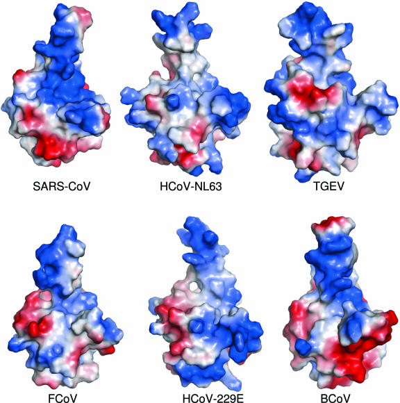
Conserved among all coronaviruses are four structural proteins: the matrix (M), small envelope (E), and spike (S) proteins that are embedded in the viral membrane and the nucleocapsid phosphoprotein (N), which exists in a ribonucleoprotein complex in the lumen. The N-terminal domain of coronaviral N proteins (N-NTD) provides a scaffold for RNA binding, while the C-terminal domain (N-CTD) mainly acts as oligomerization modules during assembly. The C terminus of the N protein anchors it to the viral membrane by associating with M protein. We characterized the structures of N-NTD from severe acute respiratory syndrome coronavirus (SARS-CoV) in two crystal forms, at 1.17 A (monoclinic) and at 1.85 A (cubic), respectively, resolved by molecular replacement using the homologous avian infectious bronchitis virus (IBV) structure. Flexible loops in the solution structure of SARS-CoV N-NTD are now shown to be well ordered around the beta-sheet core. The functionally important positively charged beta-hairpin protrudes out of the core, is oriented similarly to that in the IBV N-NTD, and is involved in crystal packing in the monoclinic form. In the cubic form, the monomers form trimeric units that stack in a helical array. Comparison of crystal packing of SARS-CoV and IBV N-NTDs suggests a common mode of RNA recognition, but they probably associate differently in vivo during the formation of the ribonucleoprotein complex. Electrostatic potential distribution on the surface of homology models of related coronaviral N-NTDs suggests that they use different modes of both RNA recognition and oligomeric assembly, perhaps explaining why their nucleocapsids have different morphologies.


Figures

FIG. 1.
FIG. 1.
(a) Organization of SARS-CoV genome. Locations of the open reading frames (ORFs) are indicated. The boundaries of the 16 nonstructural proteins (nsp1 to nsp16) that result from proteolytic processing of the replicase polyprotein (PP1ab) by PL-Protease (green) and 3CLpro (black) are marked by vertical lines. (b) Domain organization of coronaviral N proteins. The four domains labeled are as follows: SGRD, serine-glycine-arginine-rich domain; NTD, N-terminal domain; SRD, serine-rich domain; and CTD, C-terminal domain. (c) Multiple sequence alignment of NTD domains. The region for which structural coverage is provided in this study is marked by vertical lines. Hydrophobic residues are shown in yellow. Secondary structures observed for SARS-CoV N-NTD are shown above the alignment as arrows (strand) and cylinders (helix). Positively charged residues that have been implicated in RNA binding are indicated by asterisks above the sequence. The ICTV acronyms used for each viral sequence and their corresponding database accession numbers were as follows: HCoV-229E, human coronavirus 229E (NP_073556); TCoV-NC95, turkey coronavirus NC95 strain (gi32129798); BCoV-Lun, bovine coronavirus (AAL57313); HEV-VW572, porcine hemagglutinating encephalomyelitis virus (YP_459957); TGEV-Purdue, transmissible gastroenteritis virus Purdue strain (NP_058428); HCoV-NL63 human coronavirus NL63 (YP_003771); PEDV-CV777, porcine epidemic diarrhea virus CV777 strain (NP_598314); FCoV-79-1146, feline coronavirus (YP_239358); SARS-CoV-Tor2, severe acute respiratory syndrome coronavirus-Tor2 strain (AAP41047); MHV-JHM, murine hepatitis virus JHM strain (YP_209238); HCoV-OC43, human coronavirus OC43 (NP_937954); HCoV-HKU1, human coronavirus HKU-1 (YP_173242); RCoV, Rat coronavirus (AAD33104); HECoV-4408, human enteric coronavirus 4408 (AAQ67202); CCoV, canine coronavirus; ECoV-NC99, equine coronavirus NC99 (Q9DQX6); PgCoV, pigeon coronavirus (gi58416203); PCoV, puffinosis coronavirus, gi28460530; and IBV-Beaudette, avian infectious bronchitis virus (NP_040838).
FIG. 2.
FIG. 2.
(a) Structural representation of N-NTD monomer. The structure is colored from the N terminus (blue) to the C terminus (red). (b) Distribution of electrostatic potential on the surface of N-NTD. The potential distribution was calculated by using APBS module in Pymol (6). The values range from −5 kT (red) to 0 (white) and to +5 kT (blue), where k is the Boltzmann constant and T is the temperature. The orientation of the molecule is about 180° rotation along y axis of panel a. (c) The crystal structure of the monoclinic form of SARS-CoV N-NTD over the average coordinates of the NMR structure of the same domain as reported by Huang et al. (19). The four regions along the polypeptide that differ the most between the two structures are indicated by L1 to L4. Loop L1 is colored cyan for the NMR structure and blue for the crystal structure. (d) Stereo diagram showing the Cα trace of superimposed structures of SARS-CoV N-NTD and IBV N-NTD. The cubic and monoclinic forms of SARS-CoV N-NTD are shown in green and blue, respectively, while the structure from IBV is traced in red.
FIG. 3.
FIG. 3.
Crystal packing in the two crystal forms of SARS-CoV N-NTD. (a) Side-on view of three crystallographically related monomers showing stacking interactions in the monoclinic form. (b) Larger end-on view of the same crystal showing two primary modes of packing between the β-sheet cores (green and blue monomers on the left) and the protruding hairpins of adjacent monomers (yellow and brown monomers in the middle). (c and d) Three symmetry related monomers viewed along a threefold axis of the cubic crystal form (c) and a zoomed-out stereo view showing one turn along the helical axis of the cubic form (d). Equivalent trimers are labeled A, B, and C and colored green, red, and blue.
FIG. 4.
FIG. 4.
Electrostatic charge distribution on the surfaces of homology models of coronaviral N-NTDs. As in Fig. 1, the values range from −5 kT (red) to 0 (white) and to +5 kT (blue), where k is the Boltzmann constant and T is the temperature. The sequences and database accession numbers that were used as templates are the same as in Fig. 1. The boundaries of the model correspond to the regions that align with that of the SARS construct as shown in Fig. 1c.

Similar articles

Cited by

References

    1. Baric, R. S., G. W. Nelson, J. O. Fleming, R. J. Deans, J. G. Keck, N. Casteel, and S. A. Stohlman. 1988. Interactions between coronavirus nucleocapsid protein and viral RNAs: implications for viral transcription. J. Virol. 62:4280-4287. - PMC - PubMed
    1. Blicher, T., J. S. Kastrup, S. Buus, and M. Gajhede. 2005. High-resolution structure of HLA-A*1101 in complex with SARS nucleocapsid peptide. Acta Crystallogr. D Biol. Crystallogr. 61:1031-1040. - PMC - PubMed
    1. Calvo, E., D. Escors, J. A. Lopez, J. M. Gonzalez, A. Alvarez, E. Arza, and L. Enjuanes. 2005. Phosphorylation and subcellular localization of transmissible gastroenteritis virus nucleocapsid protein in infected cells. J. Gen. Virol. 86:2255-2267. - PubMed
    1. Chang, C. K., S. C. Sue, T. H. Yu, C. M. Hsieh, C. K. Tsai, Y. C. Chiang, S. J. Lee, H. H. Hsiao, W. J. Wu, C. F. Chang, and T. H. Huang. 2005. The dimer interface of the SARS coronavirus nucleocapsid protein adapts a porcine respiratory and reproductive syndrome virus-like structure. FEBS Lett. 579:5663-5668. - PMC - PubMed
    1. Davies, H. A., R. R. Dourmashkin, and R. MacNaughton. 1981. Ribonucleoprotein of avian infectious bronchitis virus. J. Gen. Virol. 53:67-74. - PubMed

Publication types

MeSH terms

Substances

LinkOut - more resources