. 2007 Apr;81(7):3574-82.
doi: 10.1128/JVI.02569-06. Epub 2007 Jan 17.

Immediate activation fails to rescue efficient human immunodeficiency virus replication in quiescent CD4+ T cells

Affiliations

Immediate activation fails to rescue efficient human immunodeficiency virus replication in quiescent CD4+ T cells

Dimitrios N Vatakis et al. J Virol. 2007 Apr.

Abstract

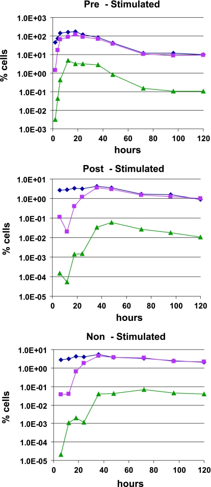
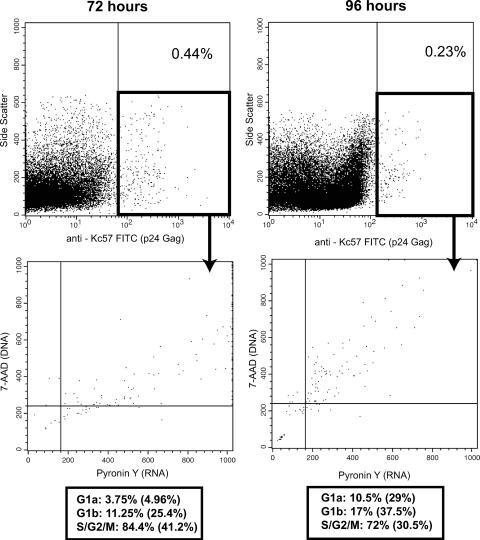
Unlike activated T cells, quiescent CD4+ T cells have shown resistance to human immunodeficiency virus (HIV) infection due to a block in the early events of the viral life cycle. To further investigate the nature of this block, we infected quiescent CD4+ T cells with HIV-1(NL4-3) and immediately stimulated them. Compared to activated (prestimulated) cells, these poststimulated cells showed slightly decreased viral entry and delays in the completion of reverse transcription. However, the relative efficiency of integration was similar to that of prestimulated cells. Together, this resulted in decreased expression of tat/rev mRNA and synthesis of viral protein. Furthermore, based on cell cycle staining and BrdU incorporation, poststimulated cells expressing viral protein failed to initiate a second round of their cell cycle, independently of Vpr-mediated arrest. Together, these data demonstrate that the early stages of the HIV life cycle are inefficient in these poststimulated cells and that efficient replication cannot be induced by subsequent activation.


Figures

FIG. 1.
FIG. 1.
Quiescent CD4+ T-cell proliferation following stimulation. Quiescent CD4 T cells were purified and stimulated as described in Materials and Methods. Cells were then harvested and stained with 7-AAD (y axis; linear fluorescence) (DNA) and Pyronin Y (x axis; linear fluorescence) (RNA) and analyzed by flow cytometry. The quadrants were set based on n-butyrate (G1a arrest) and aphidicolin (G1b arrest) treatment of the cells.
FIG. 2.
FIG. 2.
Viral entry in quiescent and prestimulated CD4+ T cells. Quiescent CD4 T cells were purified, and some were stimulated as described in Materials and Methods. Equal numbers of cells were incubated with equal amounts of virus (based on p24 values), followed by pronase treatment to remove noninternalized virus. The cells were lysed and assayed for cytosolic p24 by enzyme-linked immunosorbent assay. The data are an average of six independent experiments and indicate the levels of p24 in 1 ml of lysate. Background values of cells incubated with virus at 4°C and immediately treated with pronase were subtracted. The error bars indicate standard deviations.
FIG. 3.
FIG. 3.
Immediate activation of HIV-infected quiescent CD4+ T cells does not rescue reverse transcription and integration. Quiescent cells infected with HIV and immediately stimulated (poststimulated) or not were harvested at different times following stimulation or infection (nonstimulated groups). Prestimulated controls were also included. DNA was isolated and used as a template in a real-time PCR for the detection of initiated (⧫) and complete (▪) reverse transcripts, as well as integrated (▴) viral DNA. The levels of viral DNA in cells treated with AZT were below detection levels. For reverse transcription, 10−2% cells was the limit of sensitivity, and for the integration assays, the limit was less than 10−5%.
FIG. 4.
FIG. 4.
Viral-RNA expression is diminished in poststimulated cells. Total cellular RNA was isolated from prestimulated (diamonds), poststimulated (triangles), and nonstimulated (squares) groups and used to detect the amount of multiply spliced (tat/rev) viral RNA.
FIG. 5.
FIG. 5.
HIV gag expression is significantly decreased in poststimulated cells. Quiescent CD4+ T cells were infected with HIV at an MOI of 1 and immediately stimulated. Cells were then harvested at various times and stained with FITC-conjugated anti-Kc57 (log10 fluorescence), a Gag p24-specific antibody. No viral-protein expression was observed before 36 h poststimulation.
FIG. 6.
FIG. 6.
Gag-positive cells are mainly located in the later phases of the cell cycle. Quiescent CD4+ T cells were infected with HIV at an MOI of 1 and immediately stimulated. Cells were then harvested at various times and stained with FITC-conjugated anti-Kc57, a Gag p24-specific antibody; 7-AAD (DNA); and Pyronin Y (RNA). The distributions of the Gag-positive cells are indicated below the cell cycle plots. The percentages in parentheses refer to the total cell distribution.
FIG. 7.
FIG. 7.
Infected cells fail to complete their cell cycle. Quiescent CD4+ T cells cultured as in previous experiments in the presence of BrdU were harvested at 96 h poststimulation; stained for Gag (anti-Kc57 FITC), BrdU (anti-BrdU Alexa 647; log10 fluorescence), DNA (7-AAD), and RNA (Pyronin Y); and analyzed by flow cytometry.

References

    1. Aldrovandi, G. M., and J. A. Zack. 1996. Replication and pathogenicity of human immunodeficiency virus type 1 accessory gene mutants in SCID-hu mice. J. Virol. 70:1505-1511. - PMC - PubMed
    1. Arlen, P. A., D. G. Brooks, L. Y. Gao, D. Vatakis, H. J. Brown, and J. A. Zack. 2006. Rapid expression of human immunodeficiency virus following activation of latently infected cells. J. Virol. 80:1599-1603. - PMC - PubMed
    1. Brooks, D. G., D. H. Hamer, P. A. Arlen, L. Gao, G. Bristol, C. M. Kitchen, E. A. Berger, and J. A. Zack. 2003. Molecular characterization, reactivation, and depletion of latent HIV. Immunity 19:413-423. - PubMed
    1. Bukrinsky, M. I., T. L. Stanwick, M. P. Dempsey, and M. Stevenson. 1991. Quiescent T lymphocytes as an inducible virus reservoir in HIV-1 infection. Science 254:423-427. - PMC - PubMed
    1. Chiu, Y. L., V. B. Soros, J. F. Kreisberg, K. Stopak, W. Yonemoto, and W. C. Greene. 2005. Cellular APOBEC3G restricts HIV-1 infection in resting CD4+ T cells. Nature 435:108-114. - PubMed

Publication types