. 2007 Feb;134(4):789-99.
doi: 10.1242/dev.000380. Epub 2007 Jan 17.

Convergent extension, planar-cell-polarity signalling and initiation of mouse neural tube closure

Affiliations

Convergent extension, planar-cell-polarity signalling and initiation of mouse neural tube closure

Patricia Ybot-Gonzalez et al. Development. 2007 Feb.

Abstract

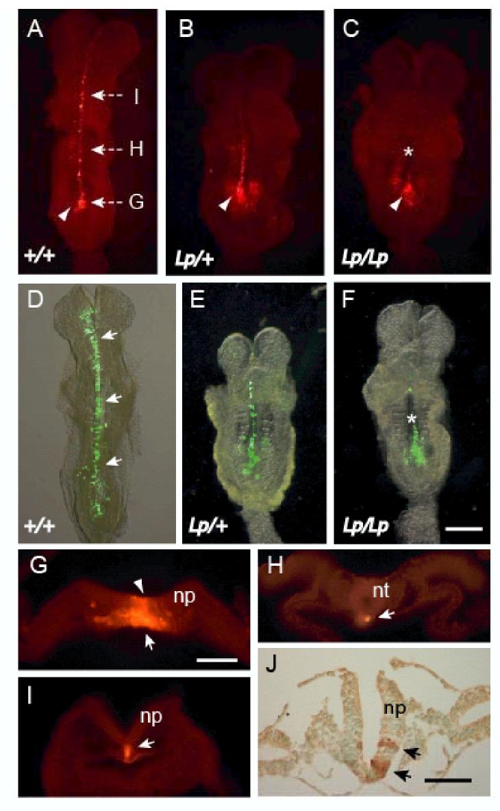
Planar-cell-polarity (PCP) signalling is necessary for initiation of neural tube closure in higher vertebrates. In mice with PCP gene mutations, a broad embryonic midline prevents the onset of neurulation through wide spacing of the neural folds. In order to evaluate the role of convergent extension in this defect, we vitally labelled the midline of loop-tail (Lp) embryos mutant for the PCP gene Vangl2. Injection of DiI into the node, and electroporation of a GFP expression vector into the midline neural plate, revealed defective convergent extension in both axial mesoderm and neuroepithelium, before the onset of neurulation. Chimeras containing both wild-type and Lp-mutant cells exhibited mainly wild-type cells in the midline neural plate and notochordal plate, consistent with a cell-autonomous disturbance of convergent extension. Inhibitor studies in whole-embryo culture demonstrated a requirement for signalling via RhoA-Rho kinase, but not jun N-terminal kinase, in convergent extension and the onset of neural tube closure. These findings identify a cell-autonomous defect of convergent extension, requiring PCP signalling via RhoA-Rho kinase, during the development of severe neural tube defects in the mouse.


Figures

Figure 1
Figure 1
Defective convergent extension in Lp embryos prior to the onset of neural tube closure. (A-C) DiI injection of the node, and (D-F) electroporation of a GFP-expressing construct into midline neural plate. Dorsal views (rostral to the top) of DiI or GFP labelled embryos, cultured for 18 h to the 5-7 somite stage. Wild type embryos (A, D) exhibit striking midline extension of DiI or GFP labelled cells (arrows), while Lp/+ embryos (B, E) exhibit variable midline extension. Lp/Lp embryos (C, F) show very limited midline extension, after either DiI or GFP labelling. Arrowheads: DiI injection site in the node; asterisks: open neural tube in Lp/Lp embryos. (G-I) Transverse sections at axial levels shown in (A) through a wild type embryo 18 h after DiI injection into the node. Caudally, intense DiI labelling is present in the notochordal plate just rostral to the node (arrow in G) and in a few midline neural plate cells (arrowhead in G). More rostrally, at the level of the closed neural tube (H) and the open hindbrain (I), only the notochord is labelled (arrows in H and I). (J) Transverse section through electroporated wild type embryo. Immunostaining with anti-GFP demonstrates labelling in neural plate only (arrows). Abbreviations: np, neural plate; nt, neural tube. Scale bars: 0.5 mm in F (also A-E); 0.1 mm in G (also H, I); 0.05 mm in J.
Figure 2
Figure 2
Diminished length and increased width of Lp/Lp embryos. (A) Embryonic length and width as measured on photomicrographs of cultured embryos viewed from the dorsal surface, after removal of yolk sac and amnion. (B-E) Length and width measurements of embryo cultured in the absence (B,C) or presence (D,E) of the ROCK inhibitor Y27632. Each data point represents a single embryo. Linear regression lines are shown. Embryonic length increases significantly with somite number in both absence (B) and presence (D) of inhibitor (2-way ANOVA; p < 0.001). Length also varies significantly with genotype (+/+ > Lp/Lp) in embryos cultured in the absence (B) of inhibitor (p = 0.008) whereas, in the presence of inhibitor (D), embryonic length no longer varies significantly with genotype (p = 0.053). Embryonic width does not vary significantly with somite number either in the absence (C) or presence (E) of inhibitor (p > 0.05). While embryonic width varies significantly between genotypes (Lp/Lp > +/+) in the absence of inhibitor (p = 0.006), there is no significant variation between genotypes in the presence of inhibitor (p > 0.05). Embryonic width is significantly greater in the presence of inhibitor (E) than in its absence (C), irrespective of genotype (p < 0.001), whereas embryonic length does not differ in the presence (D) or absence (B) of inhibitor (p > 0.05). Number of embryos measured in absence/presence of Y27632 is: +/+, 35/16; Lp/+, 56/25; Lp/Lp, 28/11.
Figure 3
Figure 3
Cell autonomous defect of convergent extension in the Lp mutant as demonstrated in chimeras. (A-C) +/++/+ and (D-F) +/+Lp/Lp chimeras at E8, produced by injecting wild type ES cells into Lp blastocysts that are also homozygous for the ROSA26 LacZ marker. After X-gal staining and eosin counterstaining, all blastocyst-derived cells are blue, while ES-derived cells are pink. Sections at the level of the notochordal plate (A,D), mid-trunk neural folds (B,E) and cranial neural folds (C,F) all show an apparently random admixture of blue and pink cells in +/++/+ chimeras (A-C) whereas +/+Lp/Lp chimeras (D-F) exhibit increased numbers of pink (wild type) cells in the midline of both neural plate (arrows) and notochordal plate (asterisk in D). More lateral regions show a more even mixture of blue and pink cells. Inset: low magnification view of +/+Lp/+ chimera showing boxed areas in which cell counts were made. (G) Quantitative analysis of relative cell numbers in midline and lateral neural plate regions of different chimera genotypes. Difference in % LacZ-negative (i.e. wild type) cells between midline and lateral regions gives values close to zero at all axial levels of +/++/+ chimeras whereas +/+Lp/Lp chimeras show values deviating markedly from zero along the entire body axis. +/+Lp/+ chimeras show a wide range of values, encompassing the extremes of the other two genotypes. Scale bar: 0.05 mm in A (also B-F).
Figure 4
Figure 4
A node defect but normal notochordal extension in E8.5 Lp embryos. (A-F) Structure of the node (arrows) in wild type (A,C,E) and Lp/Lp (B,D,F) embryos at E7.5 (A-D) and E8.5 (E,F). Embryos are homozygous for the cordon bleu (Cobl) gene trap which expresses LacZ in the node and its derivatives. At E7.5, whole mounts (A,B) and sections (C,D) show a closely similar structure and LacZ-positive cell number in the node of +/+ and Lp/Lp genotypes. In contrast, at E8.5 the LacZ-positive node appears markedly wider in sections of Lp/Lp embryos than in +/+ litter mates (E,F). (G-I) In situ hybridisation for Shh expression at E8.5 (dorsal view of whole mounts). The node (arrows) is approximately twice as wide in Lp/Lp embryos (I) as in +/+ embryos (G). Lp/+ embryos have a node of intermediate width (H). The notochordal Shh expression domain (arrowheads in G) is also broader in Lp/Lp (between white arrows in I) compared with +/+ and Lp/+. (J-L) Right lateral view of wild type E7.5 embryos a few minutes after injection of DiI into the node (J), just in front of the node (K) or further in front of the node (L). Arrows indicate position of node. (M-O) After 18 h culture, node-injected embryos exhibit labelling of the entire midline (bracketed region in M), including the triangular node/notochordal plate (white arrow in M). Injection in front of the node yields midline labelling along variable portions of the midline: either trunk + head (N), head only (O), or trunk only (not shown) depending on the precise position of injection and stage of embryo. The triangular node/notochordal plate is not labelled in these embryos. (P) Labelling in front of the node (as in K) in embryos from Lp/+ × Lp/+ litters yields a range of labelling patterns as in (N,O). The number of Lp/Lp embryos with midline extension does not differ significantly from Lp/+ and +/+ litter mates. Red dots: DiI labelled cells. Black triangles: node. Scale bars: 0.1 mm in B (also A); 0.1 mm in D (also C); 0.2 mm in E (also F); 0.2 mm in I (also G,H); 0.2 mm in L (also J,K); 0.2 mm in O (also M,N).
Figure 5
Figure 5
Expression of RhoA/ROCK and JNK signalling components during mouse neurulation. (A) RT-PCR analysis of RhoA, ROCK1, ROCK2, LIMK1 and LIMK2. Lane 1 contains molecular size standards: 800, 600 and 400 bp (top to bottom). Lanes 2-4 show expression in whole embryo homogenates at E7.5, E8.5 and E9.5 respectively. Lane 5 is no RT control. (B-J) Whole mount in situ hybridisation of wild type embryos at E8 (B-E; pre-somite headfold stage) and at E8.5 (F-J; 5-7 somites). PCP genes Vangl2 (B) and Daam1 (C), and the downstream signalling molecules RhoA (D), ROCK2 (E) are expressed throughout the late gastrulation E8 embryo. By E8.5, expression of Vangl2 (F) and Daam1 (G) becomes restricted to the hindbrain and upper spinal region, where neural tube closure is initiated (between double arrows in F-J). RhoA (H), ROCK2 (I) and JNK1 (J) are expressed in overlapping domains with the PCP genes at E8.5. Scale bars: 0.2 mm in E (also B-D); 0.2 mm in J (also F-I).
Figure 6
Figure 6
Interaction between PCP genotype and RhoA/ROCK signalling. Percent embryos with closed neural tube (i.e. successful Closure 1; upward bars) or open neural tube (i.e. failure of Closure 1; downward bars) after 18 h culture from E7.5. Data plotted against concentration of ROCK inhibitor, Y27632, or JNK inhibitor, SP600125. Hatched bars represent +/+ embryos. Solid bars represent Lp/+ (A, B), Crsh/+ (C) and Crc/+ (D) embryos. While Y27632 affects Closure 1 in wild type embryos only at the highest concentration, and not at all in circletail litters, heterozygous embryos of all three strains show failure of Closure 1 at lower Y27632 concentrations. Neurulation is not adversely affected by SP600125 in +/+ or Lp/+ embryos. For number of embryos and statistical analysis, see Table 2.
Figure 7
Figure 7
Expansion of the Shh-positive floor plate in wild type (A-H) and Lp/+ (I-L) embryos following ROCK inhibition in culture. (A) Untreated (control) wild type embryo after successful Closure 1 (arrow). (B) Wild type embryo treated with 10 μM Y27632 (Inhib), showing failed Closure 1 (arrows: entirely open neural tube). (C, D) H&E staining of transverse sections at the level of the dashed lines in A and B respectively. Note the normal, V-shaped neural fold profile in the untreated embryo (C), in contrast to the broadened floor plate region of the open neural tube after Y27632 treatment (white arrow in D). (E, F) Whole mount in situ hybridisation for Shh in wild type embryos cultured in absence (E) or presence (F) of 10 μM Y27632. Shh expression domain is broader at the site of Closure 1 (dashed lines) in the treated embryo (arrow in F) than in untreated control. (G, H) Transverse sections of Shh whole mounts, at the level of the dotted lines in E and F respectively. Shh expression domain is markedly expanded in the inhibitor-treated embryo (arrow in H). (I-L) Sections of Lp/+ embryos cultured in absence (C) or presence (D) of 2.5 μM Y27632, followed by in situ hybridisation for Shh. Expression is detected mainly in the notochord (n) of the untreated control embryo (I; higher magnification in K), with a few positive floor plate cells (white arrow in K). In the inhibitor-treated embryo (J; higher magnification in L), Shh is expressed similarly in the notochord but also in a laterally expanded floor plate domain (fp). Scale bars: 0.4 mm in F (also A, B, E), 0.1 mm in C (also D), 0.05 mm in G (also H), 0.1 mm in I (also J), 0.025 mm in K (also L).
Figure 8
Figure 8
Electroporation of constitutively active RhoA or ROCK, or wild type ROCK, disrupts convergent extension in Lp/+ and +/+ embryos. (A-F) Lp/+ and +/+ embryos after 18 h culture following electroporation in the node region with vector only (A), RhoA active (B,E), ROCK active (C,F) or ROCK wild type (D). The vector control shows marked midline extension of GFP-positive cells (arrows in A) in contrast to the sparse, clumped appearance of cells electroporated with RhoA active (arrows in B,E) or ROCK active (arrows in C,F). Electroporation with ROCK wild type allows greater midline extension (arrows in D) although less than in the vector control. (G,H) 293T cells transfected with RhoA active (H) show reduced protrusive behaviour compared with vector only controls (G). (I) Immunoblot for phosphorylated cofilin in 293T cells transfected with vector only (lane 1), RhoA active (lane 2), ROCK wild type (lane 3) or ROCK active (lane 4). Beta tubulin (lower panel) serves as a loading control. Note the severely reduced phosphocofilin abundance in cells expressing constitutively active RhoA and mild down regulation of phosphocofilin in cells expressing wild type and constitutively active ROCK. Scale bar: 0.4 mm in F (also A-E).
Figure 9
Figure 9
Summary of the convergent extension defect in Lp. Mesodermal cells emerge from the node into the notochordal plate where midline intercalation leads to narrowing to form the notochord. In the overlying neural plate, cells converge on the midline where they extend in the rostro-caudal axis. Red arrows: events that are disrupted in Lp/Lp embryos: principally midline intercalation in cells emerging from the node and convergent extension in the neural plate. Black arrows: events that appear to occur normally in Lp/Lp embryos.

Similar articles

Cited by

References

    1. Ang S-L, Rossant J. HNF-3β is essential for node and notochord formation in mouse development. Cell. 1994;78:561–574. - PubMed
    1. Bastock R, Strutt H, Strutt D. Strabismus is asymmetrically localised and binds to Prickle and Dishevelled during Drosophila planar polarity patterning. Development. 2003;130:3007–3014. - PubMed
    1. Beddington RS, Robertson EJ. An assessment of the developmental potential of embryonic stem cells in the midgestation mouse embryo. Development. 1989;105:733–737. - PubMed
    1. Beddington RSP. Induction of a second neural axis by the mouse node. Development. 1994;120:613–620. - PubMed
    1. Botto LD, Moore CA, Khoury MJ, Erickson JD. Neural-tube defects. N. Engl. J. Med. 1999;341:1509–1519. - PubMed

Publication types