. 2007 Apr;18(4):1167-78.
doi: 10.1091/mbc.e06-08-0668. Epub 2007 Jan 17.

DYRK1A autophosphorylation on serine residue 520 modulates its kinase activity via 14-3-3 binding

Affiliations

DYRK1A autophosphorylation on serine residue 520 modulates its kinase activity via 14-3-3 binding

Mónica Alvarez et al. Mol Biol Cell. 2007 Apr.

Abstract

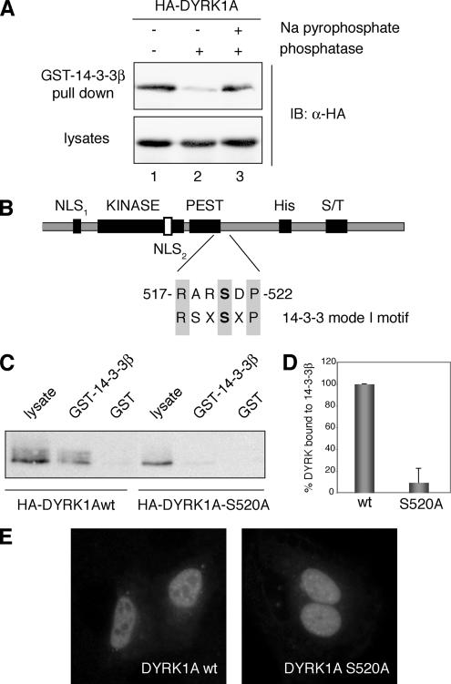
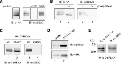
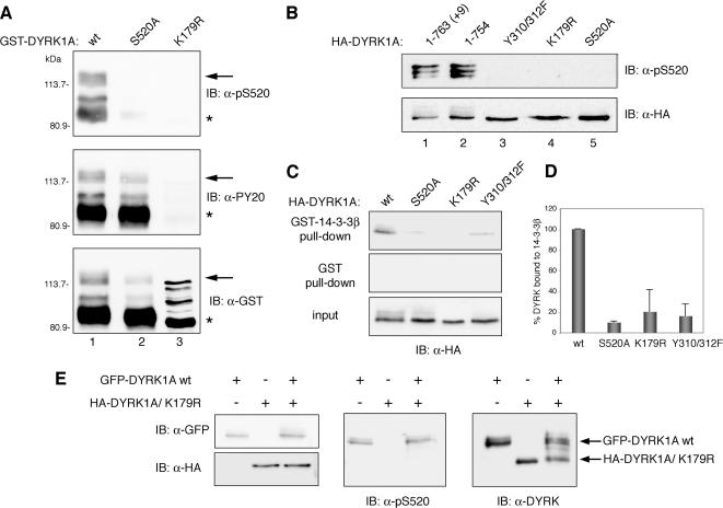
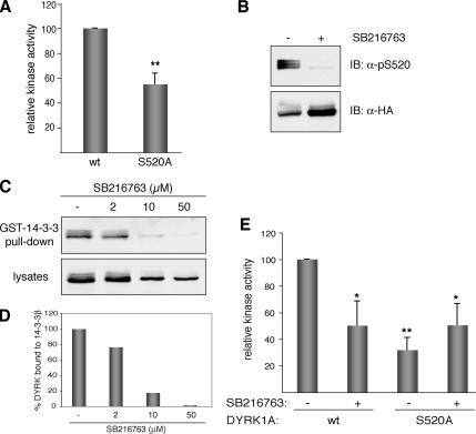
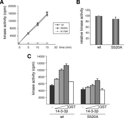
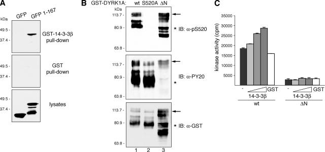
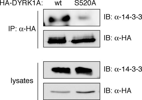
Dual-specificity tyrosine-phosphorylated and regulated kinase (DYRK) proteins are an evolutionarily conserved family of protein kinases, with members identified from yeast to humans, that participate in a variety of cellular processes. DYRKs are serine/threonine protein kinases that are activated by autophosphorylation on a tyrosine residue in the activation loop. The family member DYRK1A has been shown to phosphorylate several cytosolic proteins and a number of splicing and transcription factors, including members of the nuclear factor of activated T cells family. In the present study, we show that DYRK1A autophosphorylates, via an intramolecular mechanism, on Ser-520, in the PEST domain of the protein. We also show that phosphorylation of this residue, which we show is subjected to dynamic changes in vivo, mediates the interaction of DYRK1A with 14-3-3beta. A second 14-3-3 binding site is present within the N-terminal of the protein. In the context of the DYRK1A molecule, neither site can act independently of the other. Bacterially produced DYRK1A and the mutant DYRK1A/S520A have similar kinase activities, suggesting that Ser-520 phosphorylation does not affect the intrinsic kinase activity on its own. Instead, we demonstrate that this phosphorylation allows the binding of 14-3-3beta, which in turn stimulates the catalytic activity of DYRK1A. These findings provide evidence for a novel mechanism for the regulation of DYRK1A kinase activity.


Figures

Figure 1.
Figure 1.
Phosphorylation of Ser-520 on DYRK1A mediates its interaction with the protein 14-3-3β. (A) Lysates from HEK293 cells transiently transfected with pHA-DYRK1A were incubated for 30 min at 30°C in phosphatase buffer (lane 1) supplemented with alkaline phosphatase (lane 2) or with alkaline phosphatase and sodium pyrophosphate (lane 3) as indicated. After incubation with GST-14-3-3β immobilized on glutathione-Sepharose beads, bound protein was detected by immunoblotting (IB) with anti-HA antibody (top). An immunoblot of cell lysates representing 10% of the inputs is shown (bottom). (B) Schematic diagram of DYRK1A showing the domain structure: KINASE, kinase catalytic domain; PEST, PEST domain; His, histidine-repeat; and S/T, Ser/Thr-rich region. The sequence and the position of the putative 14-3-3 binding motif in DYRK1A are indicated and compared with a mode I 14-3-3 binding consensus sequence. The putative phosphorylated Ser residue is marked in bold. (C) HEK293 cells were transfected with pHA-DYRK1Awt and pHA-DYRK1A/S520A, and soluble lysates were incubated with unfused GST or GST-14-3-3β. Samples were analyzed by SDS-PAGE (10%) followed by immunoblotting with anti-HA antibody. Lysate represents 10% of the input. (D) The histogram presents data from three independent experiments in which the ratio bound/input in DYRK1Awt was arbitrarily set as 100. Data are expressed as a percentage of the mean value. Error bars represent SEM (standard error of the mean). (E) Subcellular localization of HA-DYRK1Awt and the mutant HA-DYRK1A/S520A, expressed in U2-OS cells, by indirect immunofluorescence analysis with anti-HA antibody followed by a FITC-conjugated anti-mouse secondary antibody.
Figure 2.
Figure 2.
DYRK1A is phosphorylated on Ser-520 in vivo. (A) Whole-cell extracts from U2-OS cells expressing HA-DYRK1Awt were resolved by 8% SDS-PAGE and analyzed by immunoblot with anti-HA antibody (left) and with anti-pS520 antibody (right). Where indicated, the phosphorylated immunizing peptide (pS520: SNSGRARpSDPTHQHR) or an equivalent unphosphorylated peptide (S520: SNSGRARSDPTHQHR) was added at 25 μg/ml during the incubation period with 1 μg/ml anti-pS520 antibody. (B) Lysates from U2-OS cells transiently transfected with pHA-DYRK1A were incubated for 30 min at 30°C in phosphatase buffer alone (lanes 1 and 3) or supplemented with alkaline phosphatase (lanes 2 and 4), as indicated. Samples were analyzed by SDS-PAGE and immunoblot with anti-HA antibody (lanes 1 and 2) and with the anti-DYRK1A pS520 antibody (lanes 3 and 4). (C) U2-OS cells were transiently transfected with pHA-DYRK1Awt (lanes 1 and 3) and pHA-DYRK1A/S520A (lanes 2 and 4). Whole-cell extracts were resolved by 8% SDS-PAGE and immunoblotted with an anti-HA antibody (lanes 1 and 2) and with the anti-DYRK1A pS520 antibody (lanes 3 and 4). (D) Soluble cell extracts from U2-OS cells expressing HA-DYRK1Awt were incubated with GST-14-3-3β. Lysates, representing 10% of the input, (lane 1) and pulled-down proteins (lane 2) were separated by 8% SDS-PAGE and analyzed by immunoblot with anti-HA antibody (bottom) and with the phospho-specific anti-DYRK1A pS520 antibody (top) to detect the presence of Ser-520–phosphorylated DYRK1A bound to 14-3-3β. (E) The endogenous DYRK1A protein was immunoprecipitated from PC12 cells with anti-DYRK1A antibody. The immunoprecipitate was analyzed by immunoblot (8% SDS-PAGE) with both the anti-DYRK1A and the anti-DYRK1A pS520 antibody. The position of marker proteins (in kilodaltons) is indicated.
Figure 3.
Figure 3.
bFGF modulate the phosphorylation status of DYRK1A Ser-520 in PC12 cells. (A) PC12 cells were serum-deprived for 20 h and then left unstimulated or stimulated with 100 ng/ml bFGF for the times indicated. Total extracts were resolved by 8% SDS-PAGE and analyzed by immunoblot on two independent membranes for the presence of phosphorylated Ser-520 and total DYRK1A. Both membranes were reprobed with anti β-tubulin antibody to confirm equal loading. Phosphorylation of the MAPK extracellular signal-regulated kinase (ERK) was detected by immunoblot with anti-phospho-p44/42 ERK (Thr202/Tyr204) antibody. (B) The chart shows the quantification of the phosphorylation levels of DYRK1A Ser-520 relative to the total amount of DYRK1A over time from the initiation of bFGF stimulation. Unstimulated cells were assigned the baseline value of 1 in each experiment. Data correspond to means ± SEM from four independent experiments. *p ≤ 0.05; **p ≤ 0.001.
Figure 4.
Figure 4.
DYRK1A autophosphorylates Ser-520 through an intramolecular reaction. (A) Bacterially expressed GST fusion proteins of wild-type DYRK1A (wt, lane 1) and the mutants DYRK1A/S520A (lane 2) and DYRK1A/K179R (lane 3) were immunoblotted with anti-DYRK1A pS520 (top), anti-phospho-Tyr (PY20; middle), and anti-GST (bottom) antibodies. The positions of marker proteins (in kilodaltons) are indicated. Arrows indicate full-length GST-fusion proteins, and asterisks indicate truncated GST-fusion proteins lacking the C-terminal (∼150) amino acids). The different patterns in DYRK1Awt, DYRK1AS520A, and DYRK1AK179R detected with the anti-GST antibody are due to differences in phosphorylation levels as shown in Supplemental Figure S3, A. (B) HA-tagged proteins corresponding to the two major isoforms of DYRK1A (lanes 1 and 2) and to distinct catalytically inactive point mutants (lanes 3–5) were overexpressed by transient transfection into U2-OS cells. Phosphorylation of Ser-520 was detected by immunoblotting with the phospho-specific anti-DYRK1A pS520 antibody (top). To control for equal expression of the fusion proteins, membranes were stripped and reprobed with an anti-HA antibody (bottom). (C) U2-OS cells were transfected with pHA-DYRK1Awt, pHA-DYRK1A/S520A, pHA-DYRK1A/K179R, or pHA-DYRK1A/Y310,312F, and soluble lysate fractions were incubated with unfused GST or GST-14-3-3β. Samples were analyzed by SDS-PAGE followed by immunoblotting with anti-HA antibody. Lysate represents 10% of input. (D) Quantification of the relative binding of different DYRK1A mutants to 14-3-3β compared with the fraction bound to DYRK1Awt is shown in the chart, which represents means ± SEM from three independent experiments. (E) A GFP-DYRK1A wild-type fusion protein (GFP-DYRK1Awt) and an HA-tagged DYRK1A kinase-inactive mutant (HA-DYRK1A/K179R) were individually expressed or coexpressed in U2-OS cells and detected by immunoblotting with anti-GFP and anti-HA antibodies, respectively (left). Phosphorylation of Ser-520 was analyzed by immunoblotting with the anti-DYRK1A pS520 antibody (middle). The expression levels of the two DYRK1A fusion proteins were compared by immunoblotting with anti-DYRK1A antibody (right).
Figure 5.
Figure 5.
Kinase activity of DYRK1A is modulated by the phosphorylation state of Ser-520. (A) US-OS cells were transfected with pHA-DYRK1Awt and the mutant pHA-DYRK1A/S520A. To determine the kinase activity of DYRK1A, tagged proteins were isolated by immunoprecipitation with anti-HA antibody, and immunocomplexes were subjected to in vitro kinase assay with DYRKtide as substrate. Equal amounts of HA-tagged proteins were used in all cases. The data represent means ± SEM from five independent experiments. **p ≤ 0.001. (B) U2-OS cells expressing HA-DYRK1Awt were treated during 8 h with the inhibitor SB216763 (50 μM) or with 0.1% DMSO as vehicle-control. Whole-cell extracts were resolved by 8% SDS-PAGE and immunoblotted with the anti-DYRK1A pS520 antibody (top). The membrane was then stripped and reprobed with anti-HA antibody to detect total HA-DYRK1A protein levels (bottom). (C) U2-OS cells expressing HA-DYRK1Awt were treated with increasing doses of SB216763, and soluble extracts were incubated with recombinant GST-14-3-3β. The amounts of bound DYRK1A (top) and DYRK1A in lysates (10% of inputs) (bottom) in each condition were analyzed by immunoblotting with anti-HA antibody. (D) The chart shows the quantification of the results expressed as the ratio of bound protein to input for each inhibitor concentration. (E) Kinase activities were determined as described in A for HA-DYRK1Awt and HA-DYRK1A/S520A expressed in U2-OS cells treated with 50 μM SB216763 for 8 h. For nontreated cells DMSO was used as vehicle-control. The data represent means ± SEM from three independent experiments. *p ≤ 0.05; **p ≤ 0.001.
Figure 6.
Figure 6.
14-3-3β binding to phospho-Ser-520 regulates DYRK1A kinase activity in vitro. (A) Kinase activity of recombinant DYRK1A proteins (GST-DYRK1Awt and the mutants S520A and K179R) was assayed with DYRKtide as substrate. The kinetics of the reaction indicated that 32P incorporation was linear with time and with the enzyme amount used in the assay. (B) The activity of GST-DYRK1Awt versus GST-DYRK1A/S520A is represented as the means ± SEM from three independent experiments, with each point measured in triplicate in the individual experiments. (C) Kinase activities of the indicated GST-DYRK1A proteins were assayed as described in A with increasing amounts of recombinant GST-14-3-3β (0.1, 0.5, and 1 μg) or GST alone (1 μg). The assay was performed three times, giving similar results in each; a representative assay is shown, and the data correspond to the means ± SEM of triplicate measurements.
Figure 7.
Figure 7.
DYRK1A harbors a second 14-3-3 binding site at the N terminus. (A) U2-OS cells were transfected with pEGFP (GFP) or pEGFP-DYRK1A/1-167 (1-167), and lysates were incubated with unfused GST or GST-14-3-3β. Samples were analyzed by immunoblotting with anti-GFP antibody. Lysates represent 10% of inputs. The positions of marker proteins (in kilodaltons) are indicated. (B) Bacterial fusion proteins corresponding to wild-type DYRK1A (wt; lane 1), the mutant DYRK1A/S520A (lane 2), and N-terminally truncated DYRK1A (ΔN; lane 3) were immunoblotted with anti-DYRK1A pS520 (top), anti-phospho-Tyr (PY20; middle), and anti-GST (bottom) antibodies. The positions of marker proteins (in kilodaltons) are indicated. Arrows indicate full-length GST-fusion proteins, and asterisks indicate truncated GST-fusion proteins lacking the C terminus. The abnormal gel mobility of the N-terminal–deleted mutant is explained by its hyperphosphorylation status as shown in Supplemental Figure S6. (C) Kinase activities of GST-DYRK1A proteins were assayed as described in Figure 6C, but GST-DYRK1Awt was compared with the N-terminally truncated mutant GST-DYRK1A/ΔN.
Figure 8.
Figure 8.
14-3-3 binding regulates DYRK1A kinase activity. (A) U2-OS cells expressing HA-DYRK1Awt and the mutant HA-DYRK1A/S520A were immunoprecipitated with anti-HA antibody. The antibody immunoprecipitated similar levels of both HA-tagged DYRK1A proteins, as detected by immunoblot with the same anti- HA antibody. The presence of endogenous 14-3-3 proteins coimmunoprecipitated with DYRK1A was detected by analyzing the samples by immunoblotting with a pan-14-3-3 antibody (top). Cells lysates represented 2% of inputs (bottom). IP, immunoprecipitation.

References

    1. Aitken A. 14-3-3 proteins: a historic overview. Semin. Cancer Biol. 2006;16:162–172. - PubMed
    1. Al-Hakim A. K., Goransson O., Deak M., Toth R., Campbell D. G., Morrice N. A., Prescott A. R., Alessi D. R. 14-3-3 cooperates with LKB1 to regulate the activity and localization of QSK and SIK. J. Cell Sci. 2005;118:5661–5673. - PubMed
    1. Altafaj X., et al. Neurodevelopmental delay, motor abnormalities and cognitive deficits in transgenic mice overexpressing Dyrk1A (minibrain), a murine model of Down's syndrome. Hum. Mol. Genet. 2001;10:1915–1923. - PubMed
    1. Alvarez M., Estivill X., de la Luna S. DYRK1A accumulates in splicing speckles through a novel targeting signal and induces speckle disassembly. J. Cell Sci. 2003;116:3099–3107. - PubMed
    1. Arron J. R., et al. NFAT dysregulation by increased dosage of DSCR1 and DYRK1A on chromosome 21. Nature. 2006;441:595–600. - PubMed

Publication types