

Pleckstrin homology (PH) domains and phosphoinositides
- PMID: 17233582
- PMCID: PMC3777418
- DOI: 10.1042/BSS0740081
Pleckstrin homology (PH) domains and phosphoinositides
Abstract
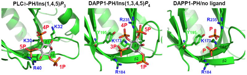
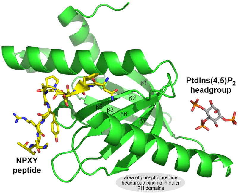
PH (pleckstrin homology) domains represent the 11th most common domain in the human proteome. They are best known for their ability to bind phosphoinositides with high affinity and specificity, although it is now clear that less than 10% of all PH domains share this property. Cases in which PH domains bind specific phosphoinositides with high affinity are restricted to those phosphoinositides that have a pair of adjacent phosphates in their inositol headgroup. Those that do not [PtdIns3P, PtdIns5P and PtdIns(3,5)P2] are instead recognized by distinct classes of domains including FYVE domains, PX (phox homology) domains, PHD (plant homeodomain) fingers and the recently identified PROPPINs (b-propellers that bind polyphosphoinositides). Of the 90% of PH domains that do not bind strongly and specifically to phosphoinositides, few are well understood. One group of PH domains appears to bind both phosphoinositides (with little specificity) and Arf (ADP-ribosylation factor) family small G-proteins, and are targeted to the Golgi apparatus where both phosphoinositides and the relevant Arfs are both present. Here, the PH domains may function as coincidence detectors. A central challenge in understanding the majority of PH domains is to establish whether the very low affinity phosphoinositide binding reported in many cases has any functional relevance. For PH domains from dynamin and from Dbl family proteins, this weak binding does appear to be functionally important, although its precise mechanistic role is unclear. In many other cases, it is quite likely that alternative binding partners are more relevant, and that the observed PH domain homology represents conservation of structural fold rather than function.
Figures
References
-
- Haslam RJ, Koide HB, Hemmings BA. Nature. 1993;363:309–310. - PubMed
-
- Mayer BJ, Ren R, Clark KL, Baltimore D. Cell. 1993;73:629–630. - PubMed
-
- Tyers M, Rachubinski RA, Stewart MI, Varrichio AM, Shorr RGL, Haslam RJ, Harley CB. Nature. 1988;333:470–473. - PubMed
-
- Yoon HS, Hajduk PJ, Petros AM, Olejniczak ET, Meadows RP, Fesik SW. Nature. 1994;369:672–675. - PubMed
-
- Macias MJ, Musacchio A, Ponstingl H, Nilges M, Saraste M, Oschkinat H. Nature. 1994;369:675–677. - PubMed
Publication types
MeSH terms
Substances
Grants and funding
LinkOut - more resources
Full Text Sources
Other Literature Sources