

Fas ligand exerts its pro-inflammatory effects via neutrophil recruitment but not activation
- PMID: 17233740
- PMCID: PMC2265864
- DOI: 10.1111/j.1365-2567.2006.02504.x
Fas ligand exerts its pro-inflammatory effects via neutrophil recruitment but not activation
Abstract
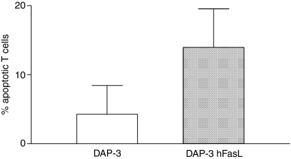
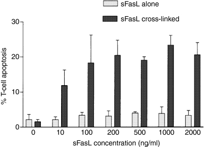
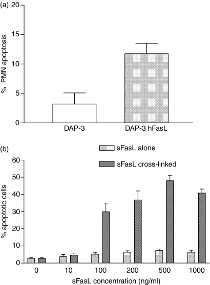
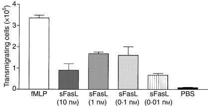
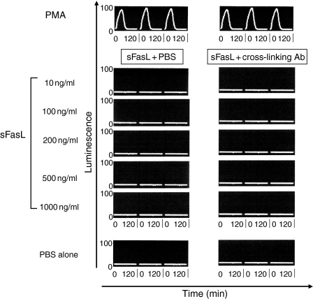
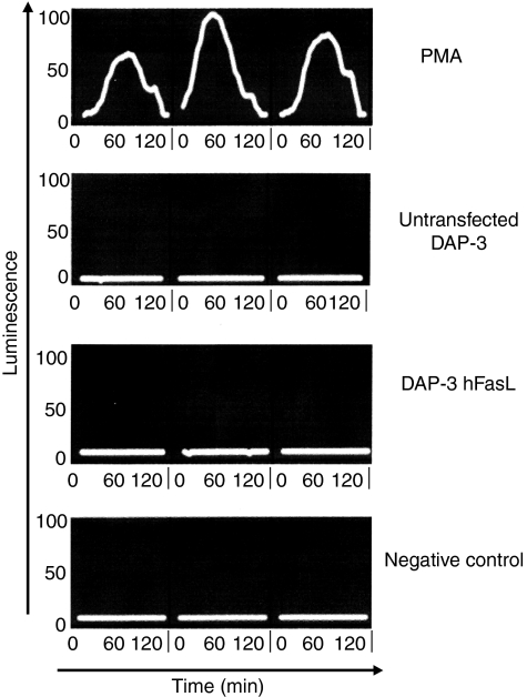
Fas ligand (FasL) expression induces apoptosis of activated T cells and has been suggested as a strategy to inhibit graft rejection. Unfortunately, the use of FasL to confer 'immune privilege' in this setting has been hampered by the finding that it may also provoke a destructive granulocytic response. While the Fas/FasL-mediated apoptotic pathways are well defined, the pro-inflammatory effects of FasL are poorly understood. Our aim in this study was to define in vitro the biological effects of FasL on neutrophil recruitment and activation. DAP-3 cells expressing human FasL on the cell membrane (mFasL) potently induced apoptosis in human neutrophils and in activated T lymphocytes. Recombinant human soluble FasL (sFasL), by contrast, was a very weak inducer of apoptosis, even at high concentrations. This latter observation suggests that cleavage of mFasL by naturally occurring matrix metalloproteinases may serve to down-regulate FasL activity in vivo. However, in the presence of a cross-linking antibody, the efficiency of apoptosis-induction by sFasL was greatly increased, suggesting that the lesser pro-apoptotic potency of sFasL reflects an inability to induce trimerization of the Fas receptor. With regard to pro-inflammatory effects, we found that sFasL is a potent neutrophil chemoattractant and, given that it induces little apoptosis, the dominance of sFasL over mFasL may mean that graft-infiltrating neutrophils will survive to mediate inflammation. Neither sFasL nor mFasL produced neutrophil activation as assessed by chemiluminescence assay. This suggests that neutrophils recruited to an inflammatory site by FasL will be activated by mechanisms other than Fas-FasL signalling.
Figures
Similar articles
-
Fas ligand is constitutively secreted by prostate cancer cells in vitro.Clin Cancer Res. 1998 Jul;4(7):1803-11. Clin Cancer Res. 1998. PMID: 9676859
-
Addressing the "Fas counterattack" controversy: blocking fas ligand expression suppresses tumor immune evasion of colon cancer in vivo.Cancer Res. 2005 Nov 1;65(21):9817-23. doi: 10.1158/0008-5472.CAN-05-1462. Cancer Res. 2005. PMID: 16267003
-
Control of ocular tumor growth and metastatic spread by soluble and membrane Fas ligand.Cancer Res. 2007 Dec 15;67(24):11951-8. doi: 10.1158/0008-5472.CAN-07-0780. Cancer Res. 2007. PMID: 18089826
-
Immune privilege or inflammation? The paradoxical effects of Fas ligand.Arch Immunol Ther Exp (Warsz). 2000;48(2):73-9. Arch Immunol Ther Exp (Warsz). 2000. PMID: 10807046 Review.
-
The role of Fas ligand and transforming growth factor beta in tumor progression: molecular mechanisms of immune privilege via Fas-mediated apoptosis and potential targets for cancer therapy.Cancer. 2004 Jun 1;100(11):2281-91. doi: 10.1002/cncr.20270. Cancer. 2004. PMID: 15160330 Review.
Cited by
-
Platelets and IgE: Shaping the Innate Immune Response in Systemic Lupus Erythematosus.Clin Rev Allergy Immunol. 2020 Apr;58(2):194-212. doi: 10.1007/s12016-019-08744-x. Clin Rev Allergy Immunol. 2020. PMID: 31254159 Review.
-
The pathological role of IL-18Rα in renal ischemia/reperfusion injury.Lab Invest. 2015 Jan;95(1):78-91. doi: 10.1038/labinvest.2014.120. Epub 2014 Oct 20. Lab Invest. 2015. PMID: 25329004
-
CD137 agonist potentiates the abscopal efficacy of nanoparticle-based photothermal therapy for melanoma.Nano Res. 2022 Mar;15(3):2300-2314. doi: 10.1007/s12274-021-3813-1. Epub 2021 Oct 12. Nano Res. 2022. PMID: 36089987 Free PMC article.
-
Interleukin-8 mediates neutrophil-endothelial interactions in pig-to-human xenogeneic models.Xenotransplantation. 2018 Mar;25(2):e12385. doi: 10.1111/xen.12385. Epub 2018 Feb 9. Xenotransplantation. 2018. PMID: 29427404 Free PMC article.
-
The microRNA miR-21 conditions the brain to protect against ischemic and traumatic injuries.Cond Med. 2017 Dec;1(1):35-46. Epub 2017 Dec 15. Cond Med. 2017. PMID: 34268484 Free PMC article.
References
-
- van Dooremal J. AkbrecgVab Graefes Arch Ophthalomol. 1873;19:358–73.
-
- Niederkorn JY. The immune privilege of corneal allografts. Transplantation. 1999;67:1503–8. - PubMed
-
- Niederkorn JY, Streilein JW, Kripke ML. Promotion of syngeneic intraocular tumor growth in mice by anterior chamber-associated immune deviation. J Natl Cancer Inst. 1983;71:193–9. - PubMed
-
- Jiang LQ, Streilein JW. Immune privilege extended to allogeneic tumor cells in the vitreous cavity. Invest Ophthalmol Vis Sci. 1991;32:224–8. - PubMed
-
- Ksander BR, Streilein JW. Immune privilege to MHC-disparate tumor grafts in the anterior chamber of the eye. I. Quantitative analysis of intraocular tumor growth and the corresponding delayed hypersensitivity response. Transplantation. 1989;47:661–7. - PubMed
Publication types
MeSH terms
Substances
LinkOut - more resources
Full Text Sources
Other Literature Sources
Research Materials
Miscellaneous
