. 2007 Mar;8(3):212-25.
doi: 10.1111/j.1600-0854.2006.00529.x.

Salmonella trafficking is defined by continuous dynamic interactions with the endolysosomal system

Affiliations

Salmonella trafficking is defined by continuous dynamic interactions with the endolysosomal system

Dan Drecktrah et al. Traffic. 2007 Mar.

Abstract

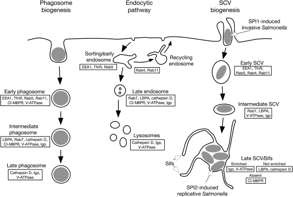
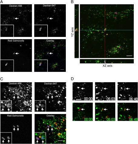
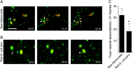
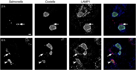
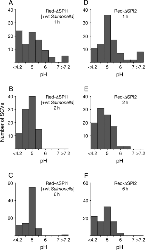
Following invasion of non-phagocytic host cells, Salmonella enterica survives and replicates within a phagosome-like compartment known as the Salmonella-containing vacuole (SCV). It is now well established that SCV biogenesis, like phagosome biogenesis, involves sequential interactions with the endocytic pathway. However, Salmonella is believed to limit these interactions and, in particular, to avoid fusion of terminal lysosomes with the SCV. In this study, we reassessed this process using a high-resolution live-cell imaging approach and found an unanticipated level of interaction between the SCV and the endocytic pathway. Direct interactions, in which late endosomal/lysosomal content was transferred to SCVs, were detected within 30 min of invasion and continued for several hours. Mechanistically, these interactions were very similar to phagosome-lysosome fusion because they were accompanied by rapid acidification of the SCV, could be blocked by chemical perturbation of microtubules or vacuolar acidification and involved the smallGTPase Rab7. In comparison with vacuoles containing internalized Escherichia coli or heat-killed Salmonella, SCVs did show some delay of fusion and acidification, although, this appeared to be independent of either type III secretion system. These results provide compelling evidence that inhibition of SCV-lysosome fusion is not the major determinant in establishment of the Salmonella replicative niche in epithelial cells.


Figures

Figure 1:
Figure 1:. Markers of the endocytic pathway and their distribution during SCV maturation
According to the prevailing model, the SCV transiently acquires markers of early endosomes and late endosomes during biogenesis, but delays delivery of the lysosomal hydrolase cathepsin D and lysobisphosphatidic acid (LBPA) and prevents the accumulation of CI-M6PRs compared to maturation of phagosomes. The SCV is also enriched in certain components of the endolysosomal pathway, particularly the V-ATPase and lgps. Markers of the late SCV also localize to membrane tubules termed Salmonella-induced filaments (Sifs).
Figure 2:
Figure 2:. SCVs acquire content from endosomes and lysosomes
To load lysosomes HeLa (A, B and D), or C2BBe1 (C), cells were preincubated with dextran-647 (red) o/n, then chased in dextran-free media for 3 h before infection with Red-Salmonella (blue). After infection, cells were incubated with dextran-488 (green) to load endosomes and live-cell imaging was initiated at 3 h p.i. A) A single confocal plane showing co-localization of dextran-647 and dextran-488 in a single SCV (arrow). B) A projection of 36 focal planes with YZ (red line) and XZ (blue line) side views to show co-localization of dextrans. C) A single confocal plane from the center of Red-Salmonella-infected C2BBe1 epithelial cells, showing co-localization of dextran-647 and dextran-488 in individual SCVs (arrows). D) Selected frames from a time-lapse series showing delivery of dextran-488 (top) from preloaded lysosomes to an SCV (arrows). Image acquisition started 15 min p.i. (00:00). Scale bars = 5 μm.
Figure 3:
Figure 3:. SCV acquisition of fluid-phase content from endosomes and lysosomes is dependent on V-ATPase and microtubules
To load lysosomes, HeLa cells were preincubated with dextran-488 o/n, then chased in dextran-free media for 3 h before infection with Red-Salmonella. To follow endocytic access to the SCV dextran-488 was added various times after infection. Cells were then observed by confocal microscopy. Images of dextran-488 and Red-Salmonella were overlaid and the number of SCVs containing dextran-488 were counted. A) Time course of SCV interaction with terminal lysosomes (filled bars) and incoming endocytic traffic (open bars). B) To assess the effect of microtubule depolymerization or V-ATPase inhibition on marker acquisition 20 μM nocodazole or 0.5 μM bafilomycin was added, respectively, 1 h before infection and maintained throughout the experiment. Live-cell imaging was initiated at 2 h p.i. and SCVs containing dextran-488 were counted. Values are the mean ± SD of three independent experiments (n ≥ 60 in each experiment). Asterisk indicates significant difference from control-treated cells (p < 0.05, anova, Tukey’s post hoc test).
Figure 4:
Figure 4:. SCV acquisition of fluid-phase markers is Rab7 dependent
HeLa cells expressing GFP alone or GFP-Rab protein fusions (green), were infected with Red-Salmonella (blue). Dextran-647 (red) was added 2 h p.i. to load the endocytic pathway. Live-cell imaging was initiated 1 h after addition of dextran-647. A single confocal plane is shown (D, overlay) to illustrate the presence of dextran-647 (B) in SCVs containing Red-Salmonella (C) that are outlined by GFP-Rab7 wt (A). Scale bar = 5 μm. E) Quantification of dextran-647 and V-ATPase positive SCVs. SCVs containing dextran-647 (gray bars) were scored in GFP-expressing cells and expressed as percentage of dextran–647 positive SCVs in mock-transfected cells (set to 100%). For quantification of V-ATPase accumulation on the SCV, transfected cells (black bars) were infected with Red-Salmonella then fixed and processed for immunofluorescence 1 h p.i. V-ATPase was detected using mouse monoclonal antibodies followed by Cy5-conjugated donkey α-mouse antibodies. SCVs staining positive for V-ATPase were counted in GFP-expressing cells and expressed as percentage of V-ATPase positive SCVs in mock-transfected cells (set to 100%). Values are the mean ± S.D. of three independent experiments (n ≥ 50 in each experiment). Asterisk indicates significant difference from all other conditions (p < 0.05, anova, Tukey’s post hoc test).
Figure 5:
Figure 5:. Increased lysosomal vesicle association with the SCV
LE/Lys were loaded by incubating HeLa cells with dextran-488 o/n, followed by a chase in dextran-free media for 3 h before infection with Red-Salmonella or Red-E. coli pInv. Live-cell imaging was initiated at 45 min p.i. and time series of 3D image volumes were collected at a rate of 28.6 frames per minute for 3.5 min. Each image volume consisted of three optical planes, spaced 0.19 μm apart, for both the dextran-488 and the Red-bacteria. Projections of selected frames are shown for Red-Salmonella (A) and Red-E. coli pInv (B) infected cells showing the association of dextran–488 containing vesicles (green, arrowheads) with vacuoles containing the bacteria (red). See Video S3 for time-lapse movies of the SCV and Video S4 E. coli pInv vacuoles. The number of vesicles detected within 1 pixel (0.19 μm) of each bacterium was quantified (C). Values are expressed as the total number of vesicles associations per bacterium in 21 frames total (every fifth frame in 100 frame series) and represent the mean ± SD of three independent experiments (n ≥ 24). Asterisk indicates significant difference p < 0.05, Student’s t-test.
Figure 6:
Figure 6:. SCVs fuse with preformed C. burnetii parasitophorous vacuoles in co-infected cells
HeLa cells were infected with C. burnetii for 48 h before infection with wt Salmonella. At 2 or 6 h p.i., cells were fixed and processed for immunofluorescence microscopy using α-Salmonella LPS (red), α-Coxiella (green) and α-LAMP1 (blue) antibodies. Projections of 12–15 confocal planes are shown. Arrows indicate Salmonella that are adjacent to, but not within, a Coxiella vacuole. An arrowhead indicates a vacuole that contains both Salmonella and Coxiella. Scale bar = 5 μm.
Figure 7:
Figure 7:. SCV acidification is delayed in epithelial cells
The endolysomal system and vacuoles containing bacteria were loaded with dextrans by incubating HeLa cells o/n and throughout infection with a mixture of dextran-FITC and dextran-647. A–D) pHSCV profiles from 0.5 to 6 h p.i. Live-cell imaging was initiated at the indicated time points after infection with Red-Salmonella. Sequential images (single optical planes) were obtained for the green (dextran-FITC), blue (dextran-647) and red (Red-Salmonella) channels. The gray scale images were combined into a single RGB image and SCVs containing Red-Salmonella and dextran-647 were selected. The fluorescence intensity ratio (488/647 nm) was acquired for each SCV and converted to pHSCV using an in situ calibration curve obtained for each sample. E) The pHVAC of HK Red-Salmonella taken up by co-internalization with wt Salmonella. F) The pHVAC of Red-E. coli pInv. (G) The pHVAC of Red-E. coli vacuoles taken up by co-internalization with wt Salmonella. The data are presented as histograms of pHVAC or pHSCV distributed in 0.5 unit bins. Each graph combines three independent experiments (n = 30 in each experiment).
Figure 8:
Figure 8:. SCVs containing ΔSPI1 or ΔSPI2 mutants do not acidify more rapidly
HeLa cells were loaded with dextran-FITC and dextran-647 as in Figure 7. A–C) Time course of the pHSCV of ΔSPI1 Red-Salmonella taken up by co-internalization with wt Salmonella. D–F) Time course of the pHSCV of ΔSPI2 Red-Salmonella internalized by SPI1-mediated invasion. The data are presented as histograms of pHSCV distributed in 0.5 unit bins. Each graph combines three independent experiments (n = 30 in each experiment).

Similar articles

Cited by

References

    1. Hueck CJ. Type III protein secretion systems in bacterial pathogens of animals and plants. Microbiol Mol Biol Rev. 1998;62:379–433. - PMC - PubMed
    1. Hensel M. Evolution of pathogenicity islands of Salmonella enterica. Int J Med Microbiol. 2004;294:95–102. - PubMed
    1. Marcus SL, Brumell JH, Pfeifer CG, Finlay BB. Salmonella pathogenicity islands: big virulence in small packages. Microbes Infect. 2000;2:145–156. - PubMed
    1. Knodler LA, Steele-Mortimer O. Taking possession: biogenesis of the Salmonella-containing vacuole. Traffic. 2003;4:587–599. - PubMed
    1. Patel JC, Galan JE. Manipulation of the host actin cytoskeleton by Salmonella– all in the name of entry. Curr Opin Microbiol. 2005;8:10–15. - PubMed

Publication types