. 2007 Jan 18:6:8.
doi: 10.1186/1476-4598-6-8.

Antiproliferative and pro-apoptotic activity of eugenol-related biphenyls on malignant melanoma cells

Affiliations

Antiproliferative and pro-apoptotic activity of eugenol-related biphenyls on malignant melanoma cells

Marina Pisano et al. Mol Cancer. .

Abstract

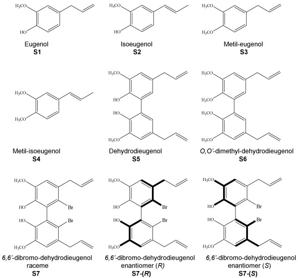
Background: Malignant melanoma is one of the most aggressive skin cancer and chemotherapeutic agents currently in use are still unsatisfactory. Prevention and early diagnosis are the only effective tools against this tumour whose incidence and mortality rates are highly increased during the last decades in fair skin populations. Therefore the search for novel therapeutic approaches is warranted. Aim of this work was to identify and test new compounds with antiproliferative and cytotoxic activity on melanoma cells. We tested eugenol together with six natural and synthetic eugenol-related compounds for their capability to inhibit cell growth on primary melanoma cell lines established from patients' tissue samples.

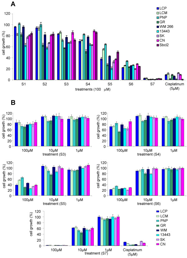
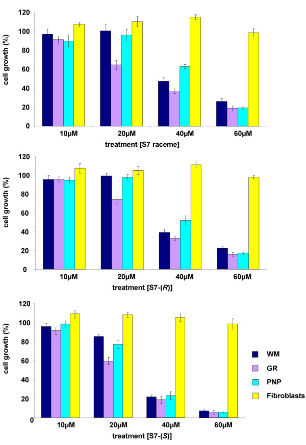
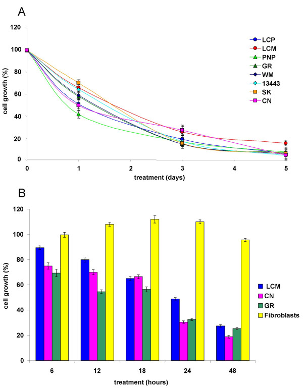
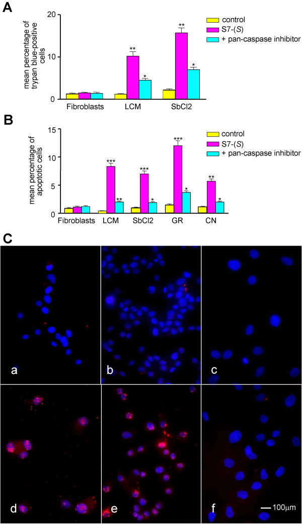
Results: Eugenol and isoeugenol monomers and their respective O-methylated forms did not show to inhibit melanoma cells proliferation. Conversely, the dimeric forms (biphenyls) showed some antiproliferative activity which was mild for dehydrodieugenol, higher for its O,O'-methylated form (O,O'-dimethyl-dehydrodieugenol), and markedly pronounced for the racemic mixture of the brominated biphenyl (6,6'-dibromo-dehydrodieugenol) (S7), being its enantiomeric form (S) the most effective compared to the other compounds. Such activity resulted to be selective against tumour cells, without affecting cultured normal human skin fibroblasts. Dose and time dependence curves have been obtained for the enantiomeric form S7-(S). Then IC50 and minimal effective doses and times have been established for the melanoma cell lines tested. TUNEL and phosphatidylserine exposure assays demonstrated the occurrence of apoptotic events associated with the antiproliferative activity of S7-(S). Cytotoxic activity and apoptosis induced by treating melanoma cells with eugenol-related biphenyls was partially dependent by caspase activation.

Conclusion: Our findings demonstrate that the eugenol related biphenyl (S)-6,6'-dibromo-dehydrodieugenol elicits specific antiproliferative activity on neuroectodermal tumour cells partially triggering apoptosis and its activity should be further investigated on in vivo melanoma models in order to evaluate the real anticancer effectiveness on such tumour.


Figures

Figure 1
Figure 1
Chemical structures of eugenol and related compounds.
Figure 2
Figure 2
Effect of eugenol derivatives on the growth of human melanoma cell lines. Nine melanoma cell lines were cultured (A) in presence of 100 μM or (B) with various concentrations of each compound (S1–S7), for 6 days and cell proliferation was estimated as described in Material and Methods. Results are expressed as percent of cell growth and represent the average (± standard deviation) of triplicate cultures performed twice. 5 μM Cisplatinum was used as cytotoxic agent positive control.
Figure 3
Figure 3
Dose-dependent S7 antiproliferative activity: comparison of raceme mixture with the enantiopure forms S7-(R) and S7-(S). Three melanoma cell lines and one short term culture of human fibroblasts were treated with various concentrations of S7, S7-(R) and S7-(S) for 3 days. Cell proliferation was estimated as described in Material and Methods. Results are expressed as percentage of cell growth and represent the average (± standard deviation) of triplicate cultures performed twice.
Figure 4
Figure 4
Time-dependent antiproliferative effect of S7-(S) on melanoma cells. (A) Eight melanoma cell lines were treated with 50 μM S7-(S) and cell proliferation was estimated at the different points, as described in Material and Methods. Results are expressed as percentage of cell growth and represent the average (± standard deviation) of triplicate cultures performed twice. (B) S7-(S) wash-out assay. Three melanoma cell lines and one short term culture of human fibroblasts were treated with 50 μM S7-(S) for various times (6–12–18–24–48 hours), then washed and incubated with S7-(S) free medium. Cell proliferation was estimated 48 hours after initiation of treatment, as described in Material and Methods. Results are expressed as percentage of cell growth and represent the average (± standard deviation) of triplicate cultures performed twice.
Figure 5
Figure 5
Effect of S7-(S) on cell viability and apoptosis in melanoma cells. (A) Melanoma cells and normal fibroblasts were incubated with 50 μM S7-(S) or solvent (control) for 24 hours, and cell death was determined by trypan blue staining. Results are expressed as mean number of trypan blue-positive cells in triplicate cultures from two independent experiments. Cells were also pre-treated for 1 hour with the pan-caspase inhibitor before S7-(S) treatment. P values for S7-(S) versus control and for inhibitor versus S7-(S) were calculated by Student's t-test with Welch correction: *, P < .05; **, P < .01. (B) Effect of S7-(S) on phosphatidylserine exposure on melanoma cells. Human fibroblasts and melanoma cell lines were incubated in the presence or absence of 50 μM S7-(S) for 24 hours. Cells were also pre-treated for 1 hour with the pan-caspase inhibitor before S7-(S) treatment. Cells were double stained with annexin V-FITC to detect phosphatidylserine exposure and propidium iodide to detect DNA and analysed by flow cytometry. Results are expressed as mean percentage of apoptotic cells from three independent experiments. P values for S7-(S) versus control and for inhibitor versus S7-(S) were calculated by Student's t-test with Welch correction: *, P < .05; **, P < .01; ***, P < .001. (C) Effect of S7-(S) on DNA fragmentation in melanoma cells. GR-mel (a, d), Sbcl2 (b, e) melanoma cell lines and human fibroblasts (c, f) were treated with 50 μM (d, e, f) S7-(S) or grown in S7-(S) free medium (control a, b, c) for 24 hours and apoptotic cells (red) were stained by the TUNEL assay. Cell nuclei (blue) were stained with DAPI.

Similar articles

Cited by

References

    1. Bataille V. Genetic epidemiology of melanoma. Eur J Cancer. 2003;39:1341–1347. doi: 10.1016/S0959-8049(03)00313-7. - DOI - PubMed
    1. Chin L. The genetics of malignant melanoma: lessons from mouse and man. Nat Rev Cancer. 2003;3:559–570. doi: 10.1038/nrc1145. - DOI - PubMed
    1. Armstrong BK, Kricker A. The epidemiology of UV induced skin cancer. J Photochem Photobiol B. 2001;63:8–18. doi: 10.1016/S1011-1344(01)00198-1. - DOI - PubMed
    1. Margolin KA. Biochemotherapy for melanoma: rational therapeutics in the search for weapons of melanoma destruction. Cancer. 2004;101:435–438. doi: 10.1002/cncr.20402. - DOI - PubMed
    1. Sondak VK, Sabel MS, Mule JJ. Allogeneic and autologous melanoma vaccines: where have we been and where are we going? Clin Cancer Res. 2006;12:2337s–2341s. doi: 10.1158/1078-0432.CCR-05-2555. - DOI - PubMed

Publication types