Comparative Study
. 2007 Jan 18:8:3.
doi: 10.1186/1471-2121-8-3.

Shared as well as distinct roles of EHD proteins revealed by biochemical and functional comparisons in mammalian cells and C. elegans

Affiliations
Comparative Study

Shared as well as distinct roles of EHD proteins revealed by biochemical and functional comparisons in mammalian cells and C. elegans

Manju George et al. BMC Cell Biol. .

Abstract

Background: The four highly homologous human EHD proteins (EHD1-4) form a distinct subfamily of the Eps15 homology domain-containing protein family and are thought to regulate endocytic recycling. Certain members of this family have been studied in different cellular contexts; however, a lack of concurrent analyses of all four proteins has impeded an appreciation of their redundant versus distinct functions.

Results: Here, we analyzed the four EHD proteins both in mammalian cells and in a cross-species complementation assay using a C. elegans mutant lacking the EHD ortholog RME-1. We show that all human EHD proteins rescue the vacuolated intestinal phenotype of C. elegans rme-1 mutant, are simultaneously expressed in a panel of mammalian cell lines and tissues tested, and variably homo- and hetero-oligomerize and colocalize with each other and Rab11, a recycling endosome marker. Small interfering RNA (siRNA) knock-down of EHD1, 2 and 4, and expression of dominant-negative EH domain deletion mutants showed that loss of EHD1 and 3 (and to a lesser extent EHD4) but not EHD2 function retarded transferrin exit from the endocytic recycling compartment. EH domain deletion mutants of EHD1 and 3 but not 2 or 4, induced a striking perinuclear clustering of co-transfected Rab11. Knock-down analyses indicated that EHD1 and 2 regulate the exit of cargo from the recycling endosome while EHD4, similar to that reported for EHD3 (Naslavsky et al. (2006) Mol. Biol. Cell 17, 163), regulates transport from the early endosome to the recycling endosome.

Conclusion: Altogether, our studies suggest that concurrently expressed human EHD proteins perform shared as well as discrete functions in the endocytic recycling pathway and lay a foundation for future studies to identify and characterize the molecular pathways involved.


Figures
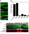
Figure 1
Figure 1
All human EHD proteins rescue the vacuolated intestinal phenotype in the intestine of C. elegans rme-1 (b1045). (A) Micrograph images of middle intestinal regions of transgenic animals expressing the human EHD proteins. The rme-1(b1045) worms were injected with pVha-6::SL2-GFP (50 ng/μl) or with the same construct containing the human EHD cDNAs along with myo2::GFP (100 ng/μl) as a co-injection marker. Intestinal vacuoles are viewed as spaces devoid of green fluorescence in the rme-1(b1045) mutant (arrows). (B) Intestinal vacuoles were counted in at least 3 independent transgenic lines expressing no vector (rme-1(b1045)), vector alone (Vector), or a vector containing EHD1-4. (C) Basolateral endocytosis assay of the intestinal vacuoles. Adult hermaphrodites were microinjected with 1 mg/mL Texas-Red BSA (TR-BSA) into the pseudocoelom and examined for uptake in intestinal vacuoles. Lack of accumulation of TRed-BSA microinjected into the pseoudocelum in wild-type (WT) worms (N2 Bristol strain) (left). Rapid accumulation of TR-BSA in the enlarged intestinal vacuoles (arrows) in the rme-1(b1045) mutant worms (middle). rme-1(b1045) worms rescued with human EHD4 do not display accumulation of the dye in any intestinal cells (right) similar to WT animals. * – pseudocoelom, ** – gonads. DIC – differential interference contrast microscopy.
Figure 2
Figure 2
All human EHD proteins are concurrently expressed in multiple cell lines. Aliquots of 100 μg cell lysates were resolved by 8% SDS-PAGE and subjected to immunoblotting with rabbit anti-peptide antisera raised against specific EHD proteins. Endogenous EHD proteins were detected in each cell lysate. The specificity of the antisera for EHD proteins is shown in Additional File 1 using GFP-fusion proteins. Relative molecular weight (MW) markers are indicated in kiloDaltons (kD). As a loading control, β-Actin was blotted.
Figure 3
Figure 3
EHD protein expression in normal mouse tissues. An 18 week-old male and female C57BL/6 mouse was sacrificed, organs were removed and lysed in tissue lysis buffer as described in Methods. Aliquots of 100 μg tissue lysate were separated using 8% SDS-PAGE and a Western blot was performed using antibodies raised against human EHD proteins. The membrane was serially stripped and reprobed beginning with both EHD1 and EHD4, followed by EHD2, EHD3 and Hsc70 antibodies. The * denotes bands that bled through from the previous blot following stripping. Differential mobility of Hsc70 may represent tissue specific isoforms. Blots shown have exposure times of less than 10 seconds, upon longer exposures, most EHD proteins can be seen in each organ shown. Relative molecular weight (MW) markers are indicated in kiloDaltons (kD). As a loading control, Hsc70 was blotted. M. gland – mammary gland.
Figure 4
Figure 4
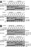
In vivo homo- and hetero-oligomer formation of human EHD proteins in mammalian cells. (A) HEK 293T cells in 100-mm tissue culture dishes were co-transfected with DNA encoding a single Myc-EHD (2.5 μg) and one EHD-GFP (2.5 μg) construct. Cell lysates were prepared 26–30 h after transfection, rocked overnight at 4°C, and 1 mg aliquots of lysate were subjected to immunoprecipitation (IP) with 3 μg of anti-Myc (9E10) antibody followed by serial anti-Myc and anti-GFP immunoblotting. A negative control (control) using 3 μg of anti-Cbl-b mouse monoclonal IgG1 (lane 9 and 19) was carried out using the lysates transfected (*) as in lane 1 and lane 11, respectively. Further negative controls for the specificity of the co-IP are shown in Additional File 2C. The identity of the lower bands in lanes 15–18 of the anti-Myc blots are unknown. (B) HEK 293T were similarly co-transfected with DNA encoding a single Myc-EHD ΔEH (2.5 μg) and an EHD-GFP (2.5 μg) construct and IPs were carried out as above except that lysates were rocked only 1–2 hours at 4°C following lysis. This exception allowed positive detection of EHD2 ΔEH co-IPs whereas EHD1 ΔEH results were unaffected with further rocking. The identity of the lower bands of the anti-Myc blots in lanes 5–9 and 15–19 are the heavy chain of the mouse IgG (IgH) and are assumed to be masked by and comigrating with Myc-EHD1 ΔEH and Myc-EHD3 ΔEH in lanes 1–4 and 11–14, respectively. All blots are representative of 3 experiments and the lysates used for these IPs are shown in Additional File 2A–2B. Relative molecular weight (MW) markers are indicated in kiloDaltons (kD).
Figure 5
Figure 5
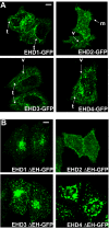
Differential subcellular localization patterns of human EHD proteins. (A) HeLa cells were transfected with C-terminal GFP-tagged EHD proteins for 24 h, fixed, mounted and scanned by a confocal microscope equipped with a 100× objective lens. Human EHD1-, 3- or 4-GFP are localized on tubular and vesicular structures in the perinuclear area, while EHD2-GFP is seen only in vesicular structures. Cells expressing EHD2 show microspikes; t, tubule; v, vesicle; m, microspikes. (B) HeLa cells were transfected with C-terminal GFP-tagged EHD ΔEH mutants for 24 h, fixed, mounted and scanned by a confocal microscope equipped with a 100× objective lens. Bar, 10 μm.
Figure 6
Figure 6
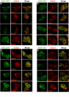
Differential colocalization of GFP- and DsRed-tagged EHD proteins co-expressed in HeLa cells. HeLa cells were co-transfected with C-terminal GFP- (green) and DsRed-tagged (red) EHD proteins for 24 h, fixed, mounted and scanned by a confocal microscope equipped with a 100× objective lens. Colocalization is indicated when similar shaped structures appear yellow in the Merge (arrowheads). (A) EHD1-GFP, (B) EHD2-GFP, (C) EHD3-GFP, (D) EHD4-GFP co-transfected with each EHD-DsRed construct. Bar, 10 μm.
Figure 7
Figure 7
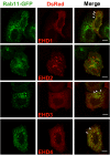
Colocalization of EHD proteins with the endocytic recycling marker Rab11. HeLa cells were co-transfected with Rab11-GFP (green) and EHD-DsRed (red) proteins for 24 h, fixed, mounted and scanned by a confocal microscope equipped with a 100× objective lens. Colocalization is indicated when similar shaped structures appear yellow in the Merge (arrowheads). Bar, 10 μm.
Figure 8
Figure 8
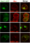
Myc-EHD1 ΔEH and EHD3 ΔEH cause perinuclear clustering of Rab11-GFP. HeLa cells were co-transfected with Myc-EHD ΔEH proteins (red) and Rab11-GFP (green) for 24 h, fixed, stained with antibodies for Myc (9E10), mounted and scanned by a confocal microscope equipped with a 100× objective lens.
Figure 9
Figure 9
Differential effects of wild type and ΔEH mutants of EHD1, 3, & 4 versus EHD2 on transferrin exit from the ERC. Untransfected HeLa cells or cells transiently transfected with Myc-EHD1 or Myc-EHD1 ΔEH were loaded with Transferrin-coupled Alexa Fluor 488 (Tf, green) in internalization buffer at 37°C for 30 minutes 24 h after transfection, washed with ice-cold PBS and chased with serum-containing medium. The cells were fixed at various time points, stained with antibodies for Myc (9E10, red) and scanned on a confocal microscope. Similar experiments with EHD2-4 and EHD2-4 ΔEH are shown in Additional File 5. (B) Cells expressing Myc-EHD proteins (black bars) or Myc-EHD ΔEH proteins (grey bars) from a representative experiment were counted with respect to Tf retention after 60 min of chase. At least 200 cells were counted in each case.
Figure 10
Figure 10
siRNA-mediated EHD protein knock-down effects on transferrin loading in HeLa cells. (A) HeLa cells were seeded on autoclaved glass coverslips in 6-well plates for 24 h followed by transfection of 200 pmol of double-stranded RNA oligonucleotides with irrelevant or EHD siRNA for 48 h prior to transferrin loading. Cells were starved for 30 min in starvation media followed by Transferrin-coupled Alexa Fluor 594 in internalization buffer at 37°C for 15 min, washed with ice-cold PBS, fixed and scanned using a confocal microscope equipped with a 40× objective lens. The arrows in the EHD1 siRNA depict an ERC transferrin loading phenotype while the arrows in the EHD4 siRNA depict an EE phenotype. (B) Cells were transfected with siRNA for EHD proteins for 24 h and further transfected with Rab5-GFP or Rab11-GFP for an additional 24 h. Cells were then loaded with labeled transferrin for 15 min as described in Methods. Arrowheads point to colocalized structures. Bar, 10 μm. Data are representative of 3 individual experiments.
Figure 11
Figure 11
siRNA-mediated EHD protein knock-down effects on transferrin recycling. Following EHD protein knock-down, transferrin loading was carried out essentially as in Figure 10 except that Transferrin-coupled Alexa Fluor 488 was loaded for 30 min. Following transferrin loading, cells were washed with ice-cold PBS, changed to serum-containing media and allowed to recycle transferrin at 37°C for various time points. The cells were fixed and scanned using a confocal microscope equipped with a 40× objective lens. Knock-down cells retaining more transferrin than irrelevant controls at 60 min of chase were considered to be delayed in recycling transferrin. Irrelevant siRNA mildly affected transferrin recycling at 30 min of chase as compared to mock. Bar, 10 μm.
Figure 12
Figure 12
Model of EHD-dependent and EHD-independent trafficking from the early and recycling endosomes. Membrane-bound receptors and their cargo (such as the transferrin receptor and transferrin, respectively) that are destined for recycling to the cell surface following endocytosis can be internalized into the early endosome (EE) compartments, transferred to the endocytic recycling compartment (ERC) before returning to the cell surface. From siRNA-mediated depletion of EHD proteins and transferrin loading and recycling experiments here and elsewhere (Naslavsky et al. 2006) [16], EHD3 and 4 are hypothesized to regulate transferrin trafficking from the EE to the ERC while EHD1 and 2 regulate transferrin exit from the ERC. Proteins such as Rabenosyn5 and Rab11-FIP2 associate with EHD proteins through EH-NPF interactions and facilitate endosomal recycling.

Similar articles

Cited by

References

    1. Mellman I. Endocytosis and molecular sorting. Annu Rev Cell Dev Biol. 1996;12:575–625. doi: 10.1146/annurev.cellbio.12.1.575. - DOI - PubMed
    1. Fazioli F, Minichiello L, Matoskova B, Wong WT, Di Fiore PP. eps15, a novel tyrosine kinase substrate, exhibits transforming activity. Mol Cell Biol. 1993;13:5814–5828. - PMC - PubMed
    1. Wong WT, Kraus MH, Carlomagno F, Zelano A, Druck T, Croce CM, Huebner K, Di Fiore PP. The human eps15 gene, encoding a tyrosine kinase substrate, is conserved in evolution and maps to 1p31-p32. Oncogene. 1994;9:1591–1597. - PubMed
    1. Confalonieri S, Di Fiore PP. The Eps15 homology (EH) domain. FEBS Lett. 2002;513:24–29. doi: 10.1016/S0014-5793(01)03241-0. - DOI - PubMed
    1. Santolini E, Salcini AE, Kay BK, Yamabhai M, Di Fiore PP. The EH network. Exp Cell Res. 1999;253:186–209. doi: 10.1006/excr.1999.4694. - DOI - PubMed

Publication types