. 2007 Mar 16;367(1):224-33.
doi: 10.1016/j.jmb.2006.12.039. Epub 2006 Dec 19.

The crystal structure of C2a, the catalytic fragment of classical pathway C3 and C5 convertase of human complement

Affiliations

The crystal structure of C2a, the catalytic fragment of classical pathway C3 and C5 convertase of human complement

Vengadesan Krishnan et al. J Mol Biol. .

Abstract

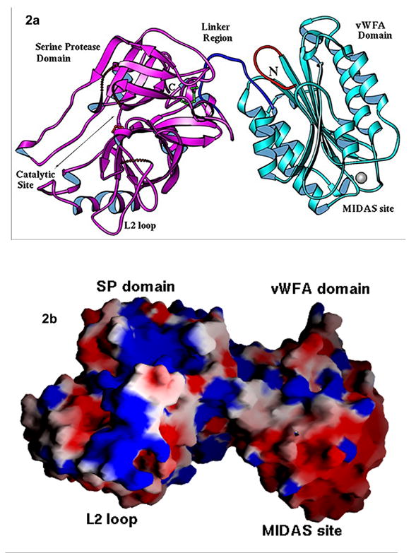
The multi-domain serine protease C2 provides the catalytic activity for the C3 and C5- convertases of the classical and lectin pathways of complement activation. Formation of these convertases requires the Mg(2+)-dependent binding of C2 to C4b, and the subsequent cleavage of C2 by C1s or MASP2, respectively. The C-terminal fragment C2a consisting of a serine protease (SP) and a von Willebrand factor type A (vWFA) domain, remains attached to C4b, forming the C3 convertase, C4b2a. Here, we present the crystal structure of Mg(2+)-bound C2a to 1.9 A resolution in comparison to its homolog Bb, the catalytic subunit of the alternative pathway C3 convertase, C3bBb. Although the overall domain arrangement of C2a is similar to Bb, there are certain structural differences. Unexpectedly, the conformation of the metal ion-dependent adhesion site and the position of the alpha7 helix of the vWFA domain indicate a co-factor-bound or open conformation. The active site of the SP domain is in a zymogen-like inactive conformation. On the basis of these structural features, we suggest a model for the initial steps of C3 convertase assembly.


Figures

Figure 1
Figure 1
a) Cartoon representation of C3-convertase formation in classical, alternative and lectin pathways of the human complement. b). Domain organization of homologous C2 and factor B of human complement. c) The SDS-PAGE of the C2 recombinant utilized in crystallization and the C2a fragment that was crystallized. From left: Lane1-LMW standards, Lane2- C2a from dissolved crystals, Lane 3- C2 protein used for crystallization after a week, Lane 4- Fresh C2 recombinant.
Figure 2
Figure 2
a). Overall ribbon representation of C2a structure. The SP domain is represented in magenta, the vWFA domain in cyan, linker in blue, and the N-terminal in red. A disulfide bond joining the linker to the SP domain and catalytic site Serine are shown in green and red respectively. b) Electrostatic surface representation of C2a. Surface charge distribution was calculated using GRASP. Positively charged regions are represented in blue, negatively charged regions in red, and both polar and nonpolar regions are in white. The marked electropositive patch represents the regions of L2 loop of SP domain
Figure 3
Figure 3
a) Overall comparison of the vWFA domain of C2a (megenta) and isolated vWFA domain of Bb (yellow). Mg ion of C2a and Bb is shown in silver and pink. The N-terminal linker of C2a is shown in red and the disordered α7 helix of Bb is shown in yellow dotted line. b) Stereo view comparison of α7 helix and its environment of C2a with other known integrin I-domain ‘open’ and ‘closed’ structures. C2 is in magenta color, ligand free α2β1 I-domain closed form is in yellow, ligand bound α2β1 I-domain is in cyan color and BbC428–C435 mutant of Bb in green color. c) Stereo view of superposition of α7 helix and its environment of C2a (magenta) and Bb (yellow). The linker to SP domain of C2a and Bb is shown in blue and green. The additional N-terminal of C2a is shown in red. The sugar molecule of C2a which is located at the domain interface near α7 helix is shown in white and labeled as NAG. Mg ion of C2a and Bb are shown in silver and yellow respectively.
Figure 4
Figure 4
(a) Stereo view of MIDAS motif comparison for ligand bound and ligand free conformations. The metal ions are colored in the same color as mains chain ribbons, and the water molecules are in blue color. The ligand free closed conformation, in the presence of Mg2+ ion for α2β1 I-domain (yellow) is superposed on ligand bound open conformation of α2β1 Id-main (cyan) and ligand free open-like conformation of C2a (magenta). A Glu residue from the ligand collagen completes the coordination of Mg in α2β1 I-domain ‘open’ conformation. (b) Stereo view of detailed comparison of MIDAS structures of C2a (magenta) and Bb (yellow). The metal atom of C2a and Bb is shown as silver and yellow ball respectively. Thin solid lines indicate the metal coordination of C2a (magenta) and Bb (yellow). Water molecules are shown in blue.
Figure 5
Figure 5
a). Overall comparison of the SP domain of C2a (megenta) and Bb (yellow). The dotted lines show the disordered loops. The catalytic triad of C2a (S659, H487, D541) and Bb (S674, H501, D551) are shown in Red and white respectively. The surface loops are labeled respectively. b). Stereo diagram showing the superposition of S1 pocket of C2a (magenta) and Bb (yellow).

Similar articles

Cited by

References

    1. Volanakis JE. Overview o the Complement System. In: Volanakis JE, Frank MM, editors. The Juman Complement System. Marcel Dekker, Inc; New York: 1998.
    1. Kamata T, Liddington RC, Takada Y. Interaction between collagen and the alpha(2) I-domain of integrin alpha(2)beta(1). Critical role of conserved residues in the metal ion-dependent adhesion site (MIDAS) region. Journal of Biological Chemistry. 1999;274:32108–11. - PubMed
    1. Arlaud GJ, Volanakis JE, Thielens NM, Narayana SV, Rossi V, Xu Y. The atypical serine proteases of the complement system. Adv Immunol. 1998;69:249–307. - PubMed
    1. Bentley DR. Primary structure of human complement component C2. Homology to two unrelated protein families. Biochem J. 1986;239:339–45. - PMC - PubMed
    1. Horiuchi T, Macon KJ, Kidd VJ, Volanakis JE. cDNA cloning and expression of human complement component C2. J Immunol. 1989;142:2105–11. - PubMed

Publication types

Substances