Comparative Study
. 2007 Jan 17;27(3):645-56.
doi: 10.1523/JNEUROSCI.4341-06.2007.

Roles of subthreshold calcium current and sodium current in spontaneous firing of mouse midbrain dopamine neurons

Affiliations
Comparative Study

Roles of subthreshold calcium current and sodium current in spontaneous firing of mouse midbrain dopamine neurons

Michelino Puopolo et al. J Neurosci. .

Abstract

We used a preparation of acutely dissociated neurons to quantify the ionic currents driving the spontaneous firing of substantia nigra pars compacta neurons, isolated from transgenic mice in which the tyrosine hydroxylase promoter drives expression of human placental alkaline phosphatase (PLAP) on the outer surface of the cell membrane. Dissociated neurons identified by fluorescent antibodies to PLAP showed firing properties similar to those of dopaminergic neurons in brain slice, including rhythmic spontaneous firing of broad action potentials and, in some cells, rhythmic oscillatory activity in the presence of tetrodotoxin (TTX). Spontaneous activity in TTX had broader, smaller spikes than normal pacemaking and was stopped by removal of external calcium. Normal pacemaking was also consistently silenced by replacement of external calcium by cobalt and was slowed by more specific calcium channel blockers. Nimodipine produced a slowing of pacemaking frequency. Pacemaking was also slowed by the P/Q-channel blocker omega-Aga-IVA, but the N-type channel blocker omega-conotoxin GVIA had no effect. In voltage-clamp experiments, using records of pacemaking as command voltage, cobalt-sensitive current and TTX-sensitive current were both sizeable at subthreshold voltages between spikes. Cobalt-sensitive current was consistently larger than TTX-sensitive current at interspike voltages from -70 to -50 mV, with TTX-sensitive current larger at voltages positive to -45 mV. These results support previous evidence for a major role of voltage-dependent calcium channels in driving pacemaking of midbrain dopamine neurons and suggest that multiple calcium channel types contribute to this function. The results also show a significant contribution of subthreshold TTX-sensitive sodium current.


Figures

Figure 1.
Figure 1.
Identification of dopaminergic neurons in midbrain of transgenic mouse expressing PLAP driven by promoter for the tyrosine hydroxylase gene. A, Coronal section of the midbrain. Both the substantia nigra and the ventral tegmental area are intensely stained by histochemical reaction for alkaline phosphatase activity reflecting presence of PLAP. Sections were incubated in 0.1% 5-bromo-4-chloro-3-indolyl phosphate and 1% nitroblue tetrazolium; histochemical procedures followed those described previously (Gustincich et al., 1997). B, Staining of neurons in the SNc by antibody to tyrosine hyroxylase. C, Higher magnification of SNc neurons after staining by reaction products caused by PLAP activity. D, Dissociated SNc neuron stained by both E6Cy3, a monoclonal antibody to PLAP directly conjugated to Cy3 (left), and by a polyclonal antibody to TH (middle). In the right panel, the images are merged. Scale bars: B, C, 50 μm; D, 15 μm.
Figure 2.
Figure 2.
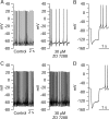
Effect of the Ih blocker ZD 7288 on pacemaking in dissociated SNc dopaminergic neurons. A, Left, Pacemaking in an acutely dissociated dopaminergic neuron from the TH-PLAP transgenic mouse line identified by Cy3-conjugated antibody to PLAP. Right, Spontaneous firing in the same cell after application of 30 μm ZD 7288. B, Response of this cell to hyperpolarizing current pulse (−80 pA current injection, before application of ZD 7228). C, Lack of effect on pacemaking of ZD 7288 applied in a different acutely dissociated neuron (also identified as dopaminergic neuron by antibody to PLAP). D, Response of this cell to hyperpolarizing current pulse (−80 pA current injection, before ZD 7288).
Figure 3.
Figure 3.
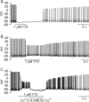
Effect of blocking calcium entry on pacemaking in a dissociated SNc dopaminergic neuron. Immediate hyperpolarization and cessation of pacemaking on switching to an external solution in which cobalt replaced calcium is evident.
Figure 4.
Figure 4.
Effect of TTX on spontaneous activity in dissociated SNc neurons. A, Hyperpolarization and cessation of pacemaking on application of TTX to a dissociated SNc neuron. B, Effect of TTX on spontaneous activity in another dissociated SNc neuron in which spontaneous activity continued, with smaller and broader spikes from a more depolarized potential. C, Effect of cobalt substitution for calcium on spontaneous activity seen in TTX in another dissociated neuron.
Figure 5.
Figure 5.
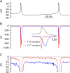
Tetrodotoxin-sensitive and cobalt-sensitive currents during the interspike interval in acutely dissociated SNc neurons determined using the action potential clamp technique. A, Spontaneous firing in a dissociated dopaminergic neuron was recorded in whole-cell current clamp. This voltage waveform was then used in the same cell as a voltage command in voltage clamp. B, Ionic currents elicited by this waveform used as command voltage after switching the amplifier to voltage clamp mode. Sodium current (red trace) was obtained by subtracting currents elicited by the voltage command before and after application of 1 μm TTX. Calcium current (blue trace) was obtained by subtracting currents elicited by the voltage command before and after replacement of calcium ions with equimolar cobalt. Inset, Sodium and calcium currents flowing during a single action potential on an expanded time scale. C, Sodium and calcium currents shown on expanded current scale. Currents were signal-averaged over 20–30 repetitions of the voltage command.
Figure 6.
Figure 6.
Comparison of current sensitive to cobalt substitution and TTX application in collected results from eight acutely dissociated SNc neurons. A, Current sensitive to TTX (red) and cobalt substitution (blue) during the interspike interval, averaged over eight neurons. Error bars show SEM. B, The contribution of cobalt-sensitive currents (blue) and TTX-sensitive sodium currents (red) during the spontaneous interspike depolarization. Currents flowing during the interspike interval were integrated from the time of maximal afterhyperpolarization to the time at which voltage reached −45 mV. In six of eight cells tested, the charge carried by calcium channels (blue bars) was bigger than the charge carried by sodium channels (red bars). Bars are stacked. Cells were arranged by their firing rate. There was no systematic correlation between firing rate and the relative contribution of sodium and calcium currents to the spontaneous interspike depolarization.
Figure 7.
Figure 7.
TTX-sensitive and cobalt-sensitive currents during the interspike interval in SNc neurons studied in brain slice. A, Spontaneous firing recorded in whole-cell current clamp in an SNc neuron in a brain slice preparation from an adult (42 d old) mouse. B, TTX-sensitive and cobalt-sensitive currents elicited in voltage clamp in the same cell using the voltage waveform from spontaneous firing as voltage command. Currents were obtained by subtracting currents before and after application of 1 μm TTX or before and after replacement of calcium ions with equimolar cobalt. C, Collected results comparing the relative contribution of TTX-sensitive and cobalt-sensitive currents during the interspike interval in eight SNc neurons studied in brain slices from 35- to 42-d-old mice. Symbols show mean ± SEM for currents measured at −65, −60, −55, and −50 mV during the interspike interval. D, Cell-by-cell comparison of charge carried by TTX-sensitive (red) and cobalt-sensitive (blue) currents during the interspike interval, measured by integrating the currents from the time of the afterhyperpolarization to the time at which voltage reached −50 mV. Cells are arranged by their firing rate, and bars are stacked. E, Comparison of mean charge carried by sodium channels (red bar) and calcium channels (blue bar) in brain slices from young (14–18 d old) animals and from older (35–42 d old) animals. Error bars show mean ± SEM for measurements from eight neurons in each age range.
Figure 8.
Figure 8.
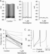
Effect of nimodipine on spontaneous firing in acutely dissociated SNc neurons. A, Spontaneous firing in an SNc neuron in control (left), after application of 1 μm nimodipine (middle), and after application of 3 μm nimodipine (right). One micromolar nimodipine produced hyperpolarization of the membrane potential (−2.1 mV) and reduction of the firing rate by ∼84%. Three micromolars nimodipine produced additional hyperpolarization and silenced the cell. B, Effect of 1 μm nimodipine on the firing rate in each of the 17 cells tested (gray symbols). In averaged results (black symbols), 1 μm nimodipine reduced firing rate by 68 ± 8% (n = 17). C, Pacemaking cycle before (black) and after (gray) exposure of a neuron to 1 μm nimodipine. Averaged pacemaking cycle was obtained by signal averaging over multiple cycles in each case, aligning spikes at their peak. Note the hyperpolarization of the membrane potential and increase in duration of the interspike interval produced by nimodipine.
Figure 9.
Figure 9.
Effect of ω-Aga-IVA on spontaneous firing in acutely dissociated SNc neurons. A, Spontaneous firing in an SNc neuron before (left) and after (right) application of 200 nm ω-Aga-IVA. ω-Aga-IVA produced a modest hyperpolarization (−2 mV) and reduced firing rate by ∼90%. B, Effect of 200 nm ω-Aga-IVA on the firing rate in each of 14 cells tested (gray symbols). In averaged results (black symbols), 200 nm ω-Aga-IVA reduced firing rate by 83 ± 8% (n = 14). C, Signal-averaged pacemaking cycle before (black) and after (gray) exposure of a neuron to 200 nm ω-Aga-IVA. Note the hyperpolarization of the membrane potential and increase in duration of the interspike interval caused by 200 nm ω-Aga-IVA.

References

    1. Astman N, Gutnick MJ, Fleidervish IA. Persistent sodium current in layer 5 neocortical neurons is primarily generated in the proximal axon. J Neurosci. 2006;26:3465–3473. - PMC - PubMed
    1. Atherton JF, Bevan MD. Ionic mechanisms underlying autonomous action potential generation in the somata and dendrites of GABAergic substantia nigra pars reticulata neurons in vitro. J Neurosci. 2005;25:8272–8281. - PMC - PubMed
    1. Banerjee SA, Hoppe P, Brilliant M, Chikaraishi DM. 5′ flanking sequences of the rat tyrosine hydroxylase gene target accurate tissue-specific, developmental, and transsynaptic expression in transgenic mice. J Neurosci. 1992;12:4460–4467. - PMC - PubMed
    1. Bennett BD, Callaway JC, Wilson CJ. Intrinsic membrane properties underlying spontaneous tonic firing in neostriatal cholinergic interneurons. J Neurosci. 2000;20:8493–8503. - PMC - PubMed
    1. Beurrier C, Bioulac B, Hammond C. Slowly inactivating sodium current (I(NaP)) underlies single-spike activity in rat subthalamic neurons. J Neurophysiol. 2000;83:1951–1957. - PubMed

Publication types

LinkOut - more resources