. 2007 Jan 30;104(5):1655-60.
doi: 10.1073/pnas.0610291104. Epub 2007 Jan 18.

An infectious retrovirus susceptible to an IFN antiviral pathway from human prostate tumors

Affiliations

An infectious retrovirus susceptible to an IFN antiviral pathway from human prostate tumors

Beihua Dong et al. Proc Natl Acad Sci U S A. .

Abstract

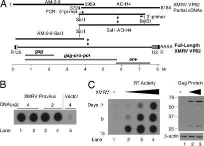
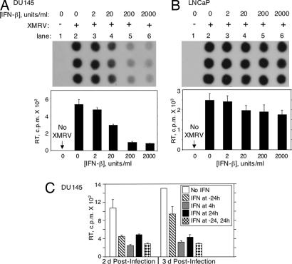
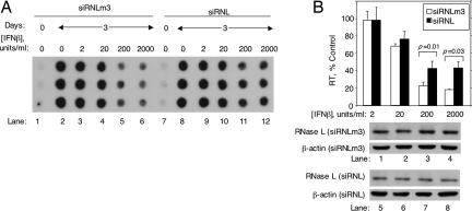
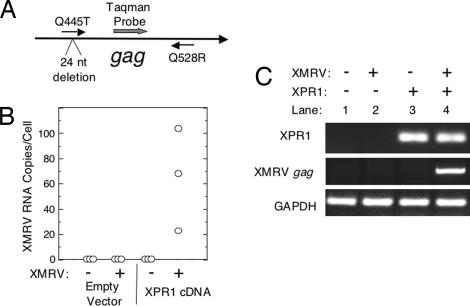
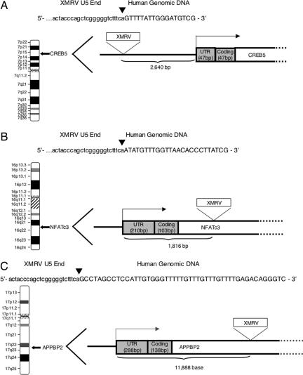
We recently reported identification of a previously undescribed gammaretrovirus genome, xenotropic murine leukemia virus-related virus (XMRV), in prostate cancer tissue from patients homozygous for a reduced activity variant of the antiviral enzyme RNase L. Here we constructed a full-length XMRV genome from prostate tissue RNA and showed that the molecular viral clone is replication-competent. XMRV replication in the prostate cancer cell line DU145 was sensitive to inhibition by IFN-beta. However, LNCaP prostate cancer cells, which are deficient in JAK1 and RNase L, were resistant to the effects of IFN-beta against XMRV. Furthermore, DU145 cells rendered deficient in RNase L with siRNA were partially resistant to IFN inhibition of XMRV. Expression in hamster cells of the xenotropic and polytropic retrovirus receptor 1 allowed these cells to be infected by XMRV. XMRV provirus integration sites were mapped in DNA isolated from human prostate tumor tissue to genes for two transcription factors (NFATc3 and CREB5) and to a gene encoding a suppressor of androgen receptor transactivation (APPBP2/PAT1/ARA67). Our studies demonstrate that XMRV is a virus that has infected humans and is susceptible to inhibition by IFN and its downstream effector, RNase L.


Conflict of interest statement

The authors declare no conflict of interest.

Figures

Fig. 1.
Fig. 1.
Cloning of full-length, replication-competent XMRV strain VP62. (A) Cloning strategy for assembling complete XMRV molecular viral clone VP62. The cloning diagram is aligned to the gene map. (B) Radiolabeled RT products from CM of LNCaP cells transfected for 10 days with 4 μg (lanes 1 and 2) and 2 μg (lanes 3 and 4) of VP62/pcDNA3.1 or from 4 μg of empty vector pcDNA3.1 (lane 5). (C Left) RT products in CM from DU145 cells previously exposed to 100 μl of CM from LNCaP cells transfected with pcDNA3.1 (lane 1) or to 1 μl (lane 2), 10 μl (lane 3), and 100 μl (lane 4) of CM from LNCaP cells transfected with VP62/pcDNA3.1. (C Right) Western blot for Gag and β-actin from DU145 cells incubated for 9 days with 100 μl of CM from pcDNA3.1-transfected LNCaP cells (lane 1) or CM from VP62/pcDNA3.1-transfected LNCaP cells. Lane 2, 10 μl of CM; lane 3, 100 μl of CM.
Fig. 2.
Fig. 2.
IFN sensitivity of XMRV in DU145 and LNCaP cells. (A and B) DU145 cells (A) or LNCaP cells (B) plated and assayed in triplicate were incubated for 16 h in the absence or presence of different amounts of IFN-β as indicated and then mock-infected (lane 1) or infected with XMRV (lanes 2–6) for 3 days. IFN-β was added a second time at 24 h after infection. (Upper) Autoradiograms of the radiolabeled RT products. (Lower) RT activity (cpm) as a function of [IFN-β]. (C) Effect of time of IFN-β treatment on viral yields in DU145 cells. The IFN added at −24 h only was removed and not replaced at the time of infection. Assays were performed in triplicate. The decreases in RT activity in response to IFN treatments were significant. P = 0.014 and 0.018 in two-tailed, paired Student's t tests at 20 and 2,000 units/ml IFN-β in A and B, respectively.
Fig. 3.
Fig. 3.
Effect of RNase L on the antiviral activity of IFN-β. (A and B Top) RT activities from CM of DU145 cells expressing short hairpin RNA to RNase L (siRNL) or expressing a three-base mismatch control RNA (siRNLm3) (as indicated). Cells were infected for 12 days with XMRV before addition of IFN-β. Lanes 1 and 7, media control; lanes 2–6 and 8–12, CM after 3 days of IFN treatment. (B Middle and Bottom) Western blots for RNase L and β-actin were from the same experiment, blot, and exposure. Two-tailed, paired Student's t tests were performed in B.
Fig. 4.
Fig. 4.
Infection of hamster cells with XMRV depends on expression of human XPR1. (A) Diagram of quantitative real-time RT-PCR strategy for amplifying an 84-bp region from the 5′ UTR of XMRV gag (nucleotides 445–528). (B) XMRV RNA copy number in CHO cells transiently transfected or mock-infected with empty vector pcDNA3.1 or human XPR1 cDNA in vector pcDNA3.1 followed by exposure to XMRV and continuous culturing for 30 days. The experiment was performed in triplicate. (C) Nested RT-PCR for XPR1, XMRV gag, and GAPDH RNAs in a representative experiment from B. An agarose gel in which the PCR products were stained with ethidium bromide is shown.
Fig. 5.
Fig. 5.
Locations of XMRV integration sites in prostate DNA from case VP268 (A and B) and case VP234 (C). Genomic DNA was isolated from the patient tumor sample, and the DNA sequence near the virus–host DNA junction was cloned and sequenced (Materials and Methods). (A) In chromosome 7p15.1 the integrated provirus was 2,640 bp upstream of the CREB5 transcription start site. (B) In chromosome 16q22.1 the integrated provirus was 1,816 bp downstream of the NFATc3 transcription start site. (C) In chromosome 17q23.2 the integrated provirus was 11,888 bp downstream of the APPBP2 transcription start site. Lowercase letters represent the sequence at the U5 end of the viral long terminal repeat, and uppercase letters represent human genomic sequences. Arrows denote the virus–host DNA junctions. Right-angled arrows denote the transcription start sites.

Comment in

Similar articles

Cited by

References

    1. Goff S. In: Fields Virology. Knipe DM, Howley PM, editors. New York: Lippincott Williams & Wilkins; 2001. pp. 1871–1939.
    1. Bartosch B, Weiss RA, Takeuchi Y. J Gen Virol. 2002;83:2231–2240. - PubMed
    1. Hanger JJ, Bromham LD, McKee JJ, O'Brien TM, Robinson WF. J Virol. 2000;74:4264–4272. - PMC - PubMed
    1. Urisman A, Molinaro RJ, Fischer N, Plummer SJ, Casey G, Klein EA, Malathi K, Magi-Galluzzi C, Tubbs RR, Ganem D, et al. PLoS Pathog. 2006;2:e25. - PMC - PubMed
    1. Carter BS, Bova GS, Beaty TH, Steinberg GD, Childs B, Isaacs WB, Walsh PC. J Urol. 1993;150:797–802. - PubMed

Publication types

Associated data