. 2007 Jan 15;21(2):160-74.
doi: 10.1101/gad.1503107.

The Spt6 SH2 domain binds Ser2-P RNAPII to direct Iws1-dependent mRNA splicing and export

Affiliations

The Spt6 SH2 domain binds Ser2-P RNAPII to direct Iws1-dependent mRNA splicing and export

Sunnie M Yoh et al. Genes Dev. .

Abstract

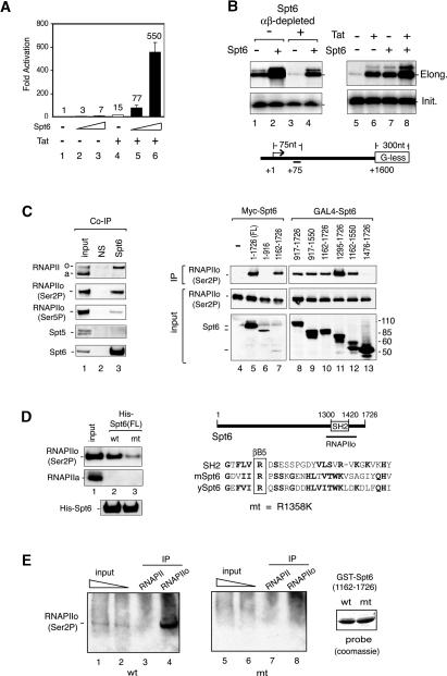
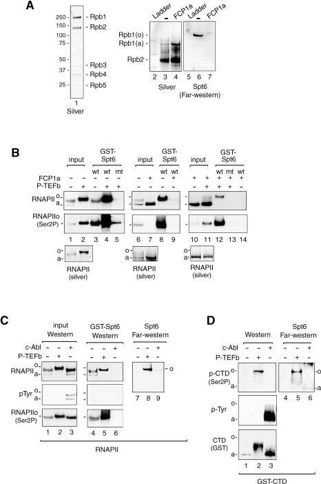
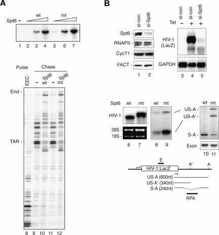
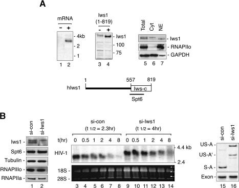
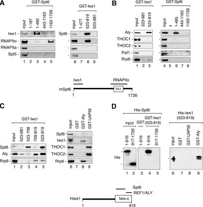
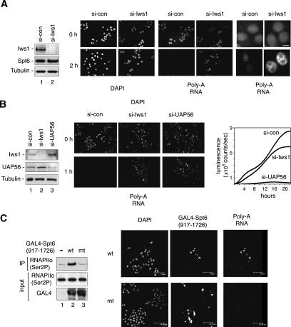
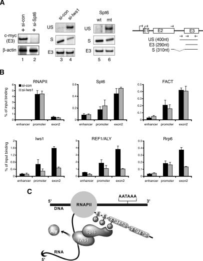
Spt6 promotes transcription elongation at many genes and functions as a histone H3 chaperone to alter chromatin structure during transcription. We show here that mammalian Spt6 binds Ser2-phosphorylated (Ser2P) RNA polymerase II (RNAPII) through a primitive SH2 domain, which recognizes phosphoserine rather than phosphotyrosine residues. Surprisingly, a point mutation in the Spt6 SH2 domain (R1358K) blocked binding to RNAPIIo without affecting transcription elongation rates in vitro. However, HIV-1 and c-myc RNAs formed in cells expressing the mutant Spt6 protein were longer than normal and contained splicing defects. Ectopic expression of the wild-type, but not mutant, Spt6 SH2 domain, caused bulk poly(A)+ RNAs to be retained in the nucleus, further suggesting a widespread role for Spt6 in mRNA processing or assembly of export-competent mRNP particles. We cloned the human Spt6-interacting protein, hIws1 (interacts with Spt6), and found that it associates with the nuclear RNA export factor, REF1/Aly. Depletion of endogenous hIws1 resulted in mRNA processing defects, lower levels of REF1/Aly at the c-myc gene, and nuclear retention of bulk HeLa poly(A)+ RNAs in vivo. Thus binding of Spt6 to Ser2-P RNAPII provides a cotranscriptional mechanism to recruit Iws1, REF1/Aly, and associated mRNA processing, surveillance, and export factors to responsive genes.


Figures

Figure 1.
Figure 1.
Spt6 binds tightly to Ser2-P RNAPII. (A) Spt6 enhances Tat transactivation of an integrated HIV-1:LacZ reporter gene in HeLa P4 cells. HeLa P4 cells were transfected with 5 or 25 ng of Spt6 vector in the presence or absence of recombinant HIV-1 Tat101, which was introduced to the cells by chloroquine-mediated protein transduction. (B) In vitro transcription analysis of an HIV-1 G-less template in untreated (lanes 1,2,5–8) or Spt6-immunodepleted (lanes 3,4) nuclear extracts in the presence of recombinant Spt6 (lanes 2,4,7), HIV-1 Tat (lane 6), or both proteins (lane 8). In vitro synthesized RNA was monitored by run-off (elongation, top panels) or primer extension (initiation, bottom panels) assays. (C) Spt6 associates with Ser2-P RNAPII in nuclear extracts. Immunoblot analysis of HeLa nuclear extract (input, lane 1), or fractions immunoprecipitated with antisera to Spt6 (lane 3) or with nonspecific IgG (NS) antisera (lane 2). Antibodies used for immunoblot are indicated to the left of each panel. (Top, Lanes 4–13, top) To map the RNAPII-interacting domain, transiently expressed Myc-tagged Spt6 or GAL4-Spt6 proteins were analyzed for associated Ser2-P RNAPII by immunoblot. (Lanes 4–13) Input RNAPIIo levels are shown in the middle panels, and the different Spt6 protein domains are shown in the bottom panels. (D, lanes 1–3) Full-length wild-type or R1358K recombinant His-Spt6 proteins (shown in the bottom panel) were coupled to beads, incubated with HeLa nuclear extract, and analyzed for binding to Ser2P RNAPII or RNAPIIa. A schematic shows the sequence of the murine Spt6 SH2 domain and the location of the R1358K mutation. (E) Far-Western analysis of binding of wild-type (wt) or R1358K mutant (mt) GST-Spt6 proteins (amino acids 1162–1726) to immunoprecipitated HeLa Ser2-P RNAPII (H5, lanes 4,8) or total RNAPII (N20, lanes 3,7). Binding of the GST-Spt6 proteins to the immunoprecipitated Ser2-P or total RNAPII complexes was visualized by incubating the membrane with glutathione-horseradish peroxidase (Glut-HRP). The panel at the right shows a Coomassie stain of the wild-type and mutant GST-Spt6 proteins used to probe the blot.
Figure 2.
Figure 2.
An SH2 domain mediates binding of Spt6 to Ser2-P RNAPII. (A) Purified HeLa RNAPII (lane 1) was incubated in the presence or absence of the FCP1a CTD phosphatase and analyzed by silver stain (lanes 2–4) or Far-Western blot (lanes 5–7). Binding of the GST-Spt6 SH2 domain (amino acids1162–1726) was detected by staining with Glut-HRP. (B) Binding of the GST-Spt6 SH2 domain (amino acids 1162–1496) to purified HeLa RNAPII was analyzed in the absence or presence of the FCP1a CTD phosphatase or P-TEFb CTD kinase, as indicated above each lane. The RNAPII complexes bound to the GST-Spt6 beads were identified by immunoblot using antisera to total (top) or Ser2-P (middle) RNAPII, and the input RNAPII fractions were also analyzed by silver stain (bottom). (C) Binding of the GST-Spt6 SH2 domain (amino acids 1162–1496) to P-TEFb-phosphorylated (lanes 2,5,8) or c-Abl-phosphorylated (lanes 3,6,9) HeLa RNAPII complexes was examined by means of GST-pulldown (lanes 4–6) or Far-Western blot (lanes 7–9) experiments. Interactions were visualized with antisera to total RNAPII (top), phosphotyrosine (pTyr, middle), or Ser2-P RNAPII (bottom), as indicated next to each panel. The Far-Western blot was visualized with Glut-HRP stain. (D) Far-Western analysis of binding of the GST-Spt6 SH2 domain (amino acids 1162–1726) to purified recombinant murine GST-CTD, either alone (lanes 1,4), or after incubation with recombinant P-TEFb (lanes 2,5) or c-Abl (lanes 3,6) kinases.
Figure 3.
Figure 3.
Spt6 binds Ser2-P RNAPII to regulate mRNA processing, but not RNAPII transcription elongation. (A, lanes 1–7) Purified recombinant wild-type and R1358K Spt6 proteins enhance RNAPII transcription elongation in vitro. Reactions contained 10 ng (lanes 2,5), 25 ng (lanes 3,6), or 50 ng (lanes 4,7) of wild-type (lanes 2–4) or mutant (lanes 5–7) Spt6 protein. The rate of transcription elongation by recombinant full-length wild-type (lane 10) or R1358K mutant Spt6 (lane 12) was analyzed by pulse-chase analysis of isolated EECs (lane 8). (Lanes 9,11) Where indicated, the HIV-1 EEC was incubated with 100 ng of wild-type Spt6, R1358K Spt6, or GST control, and transcripts were extended following a chase with excess ribonucleotides. (B) siRNA-mediated depletion of endogenous Spt6 blocks transcription of the integrated HIV-1 reporter gene in HeLa P4 cells. Cells treated with control (si-con) or Spt6 (si-Spt6) siRNAs for 84 h were analyzed by immunoblot (lanes 1,2), or by Northern blot for expression of HIV-1:LacZ or GAPDH RNA (lanes 3–5). The bottom panels show Northern blot analyses of HIV-1:LacZ transcripts in HeLa P4 cells expressing siRNA-resistant wild-type (lane 6) or R1358K mutant (lane 7) Spt6 proteins. In this experiment, cells were first treated with an Spt6-siRNA to deplete endogenous Spt6, and HIV-1 transcription was induced by cotransfection with an HIV-1 Tat expression construct. Levels of spliced and unspliced HIV-1:LacZ transcripts were assessed by RPA (lanes 8,9) and RT–PCR (lanes 10,11). (Bottom panel) Quantitative RT–PCR using primers within the exon in HIV-1:LacZ served as an RNA input control. The schematic diagram depicts the integrated HIV-1:LacZ gene in HeLa P4 cells. The HIV-1:LacZ transcripts were amplified using LacZ gene-specific (arrow) and a oligo-dT anchor primers. (S) Spliced transcripts; (US) unspliced transcripts; (A) major polyadenylation sites; (A′) premature polyadenylation sites; (E) Northern probes; (RPA) RPA probes.
Figure 4.
Figure 4.
The human Iws1 protein is necessary for splicing of HIV-1:LacZ mRNA in vivo. (A) Analysis of human Iws1 gene expression by Northern blot of HeLa mRNA (lanes 1,2) and immunoblot (lanes 3,4) of a lysate from cells transfected with the hIws1 cDNA (lane 4). (Lanes 5–7) Nuclear and cytoplasmic HeLa extracts were analyzed for hIws1 by immunoblot. At the bottom is a schematic of the hIws1 protein, indicating a C-terminal region (Iws-C, boxed) that is homologous to the S. cerevisiae Iws1/SPN1 protein. (B) Northern blot analysis of HIV-1:LacZ RNA in cells transfected with control- or Iws1-specific siRNAs. (Lanes 1,2) Specific depletion of endogenous hIws1 was assessed by immunoblot. (Lanes 3–14) Cells were treated with ActD (5 μg/mL) for the various times indicated above each lane to block ongoing transcription, and HIV-1:LacZ transcripts were analyzed by Northern blot. (Lanes 15,16) RT–PCR analysis of HIV-1:LacZ RNA in the absence of ActD (0 h) in cells treated with control or Iws1-siRNAs.
Figure 5.
Figure 5.
Human Iws1 interacts directly with Spt6 and REF1/Aly in vitro. (A) GST-Spt6 (lanes 1–5) or GST-Iws1 (lanes 6–9) proteins were coupled to beads, incubated with HeLa nuclear extract, and analyzed for binding to RNAPII and other factors by immunoblot using the antisera indicated to the left of each panel. (B) Immunoblot analysis of the REF1/Aly and other factors to GST-Iws (lanes 1–3) or GST-Spt6 (lanes 4–8) protein-coupled beads in GST-pulldown experiments with HeLa nuclear extract. (C) Analysis of the ability of different transcription factors to bind GST-hIws1 domains (lanes 1–5), GST (lane 7), GST-Aly (lane 8), or GST-UAP56 (lane 9) in GST-pulldown experiments carried out with HeLa nuclear extract. Interacting proteins were visualized by immunoblot using the antisera indicated to the left of each panel. (D) Direct binding of mSpt6 to hIws1 and of hIws1 to hREF1/Aly was analyzed in GST-pulldown experiments carried out with purified recombinant factors. Recombinant His-Spt6 (amino acids 1–916 or 917–1726) proteins were incubated with GST (lane 3) or GST-Iws1 (amino acids 523–819; lanes 4,5) beads, and binding was assessed by immunoblot. For the right panel, His-Iws1 (amino acids 523–819) protein was incubated with GST (lane 7), GST-UAP56 (lane 8), and GST-Aly (lane 9) beads, and binding was examined by immunoblot.
Figure 6.
Figure 6.
Depletion of endogenous hIws1 impairs export of bulk polyadenylated mRNAs. (A) HeLa cells were transfected with control or hIws1-siRNAs and analyzed by immunoblot (lanes 1,2), immunofluorescence microscopy (DAPI stain), or RNA-FISH to detect poly(A)+ RNAs, as indicated. Cells were either untreated (top panels) or treated with ActD (5 μg/mL) for 2 h (bottom panels) prior to RNA-FISH. The panels on the far right show the fixed nuclei at higher magnification. Bar, 10 μm. (B) HeLa cells were transfected with si-control, si-Iws1, or si-UAP56, and analyzed by immunoblot (lanes 1–3) or by RNA-FISH for poly(A)+ RNAs (bottom panels). Cells were treated with ActD for 1 h prior to RNA-FISH. Graph at the far right compares the rate of expression of an HIV-1:luciferase reporter gene in Tat-expressing HeLa cells treated with control-, Iws1-, or UAP56-specific siRNAs. Luciferase levels were measured by luminescence in counts per second over a 22-h time period. (C) Overexpression of the GAL4-Spt6 SH2 domain blocks the nuclear export of poly(A)+ RNAs in HeLa cells. (Lanes 1–3) Transiently expressed wild-type or mutant GAL4-Spt6 SH2 domain proteins were immunoprecipitated with an anti-GAL4 antibody, and binding to RNAPIIo was determined by immunoblot. The panels show RNA-FISH analysis of HeLa cells transfected with wild-type (wt) or R1358K mutant (mt) GAL4-Spt6-SH2 (amino acids 917–1726) domain proteins. Cells were treated with ActD for 1 h and analyzed by DAPI stain, immunostaining with GAL4-specific antisera, or in situ hybridization for poly(A)+ RNA, as indicated above each panel. Bar, 75 μm.
Figure 7.
Figure 7.
The hIws1 protein recruits REF1/Aly to the c-myc gene in vivo. (A) RT–PCR analysis of c-myc mRNA levels in 293T cells expressing control siRNA (lanes 1,3), Spt6-siRNA (lane 2), hIws1-siRNA (lane 4), or the mutant R1358K Spt6 protein (lane 6). The schematic shows RT–PCR products of spliced (S), unspliced (US), and total (E3) c-myc mRNA. (B) ChIP analysis of the c-myc gene in 293T cells treated with control or hIws1-siRNAs. Chromatin was immunoprecipitated with the indicated antisera and analyzed by Q-PCR using primers specific for the enhancer, promoter, or second exon of the human c-myc gene. (C) Working model depicting the association of hIws1 with Spt6 bound to the Ser2-P RNAPII CTD. Our data indicate that this complex is required to facilitate steps required for proper mRNA processing and export, and may also serve to recruit REF1/Aly to Spt6-dependent genes.

References

    1. Abruzzi K.C., Lacadie S., Rosbash M., Lacadie S., Rosbash M., Rosbash M. Biochemical analysis of TREX complex recruitment to intronless and intron-containing yeast genes. EMBO J. 2004;23:2620–2631. - PMC - PubMed
    1. Adelman K., Wei W., Ardehali M.B., Werner J., Zhu B., Reinberg D., Lis J.T., Wei W., Ardehali M.B., Werner J., Zhu B., Reinberg D., Lis J.T., Ardehali M.B., Werner J., Zhu B., Reinberg D., Lis J.T., Werner J., Zhu B., Reinberg D., Lis J.T., Zhu B., Reinberg D., Lis J.T., Reinberg D., Lis J.T., Lis J.T. Drosophila Paf1 modulates chromatin structure at actively transcribed genes. Mol. Cell. Biol. 2006;26:250–260. - PMC - PubMed
    1. Adkins M.W., Tyler J.K., Tyler J.K. Transcriptional activators are dispensable for transcription in the absence of Spt6-mediated chromatin reassembly of promoter regions. Mol. Cell. 2006;21:405–416. - PubMed
    1. Aguilera A. Cotranscriptional mRNP assembly: From the DNA to the nuclear pore. Curr. Opin. Cell Biol. 2005;17:242–250. - PubMed
    1. Ahn S.H., Kim M., Buratowski S., Kim M., Buratowski S., Buratowski S. Phosphorylation of serine 2 within the RNA polymerase II C-terminal domain couples transcription and 3′ end processing. Mol. Cell. 2004;13:67–76. - PubMed

Publication types

MeSH terms