. 2007 May;25(5):1126-35.
doi: 10.1634/stemcells.2006-0540. Epub 2007 Jan 18.

Selection of embryonic stem cell-derived enhanced green fluorescent protein-positive dopamine neurons using the tyrosine hydroxylase promoter is confounded by reporter gene expression in immature cell populations

Affiliations

Selection of embryonic stem cell-derived enhanced green fluorescent protein-positive dopamine neurons using the tyrosine hydroxylase promoter is confounded by reporter gene expression in immature cell populations

Eva Hedlund et al. Stem Cells. 2007 May.

Abstract

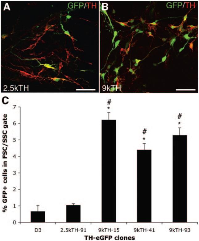
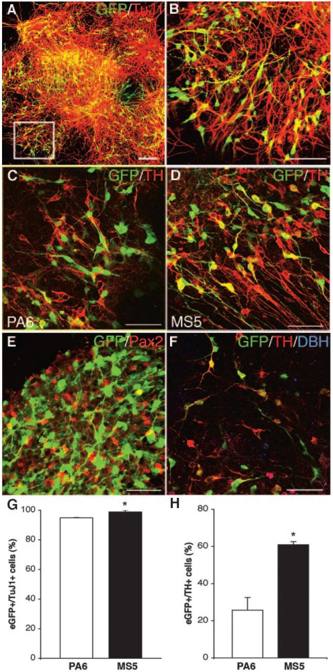
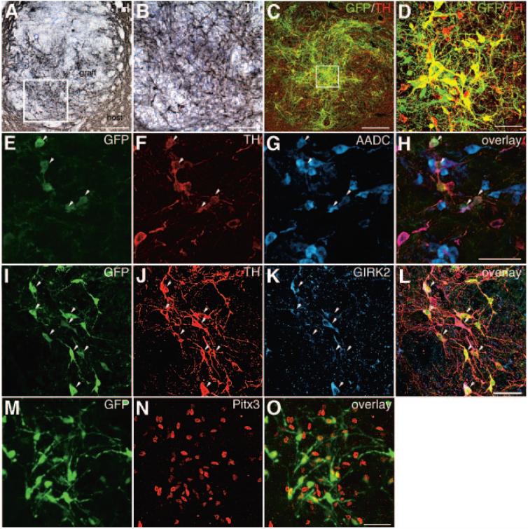
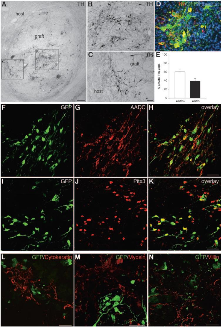
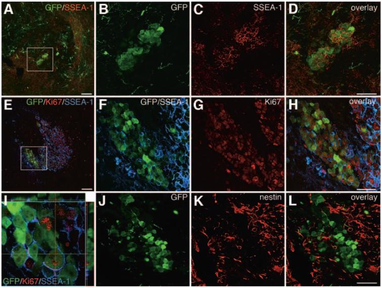
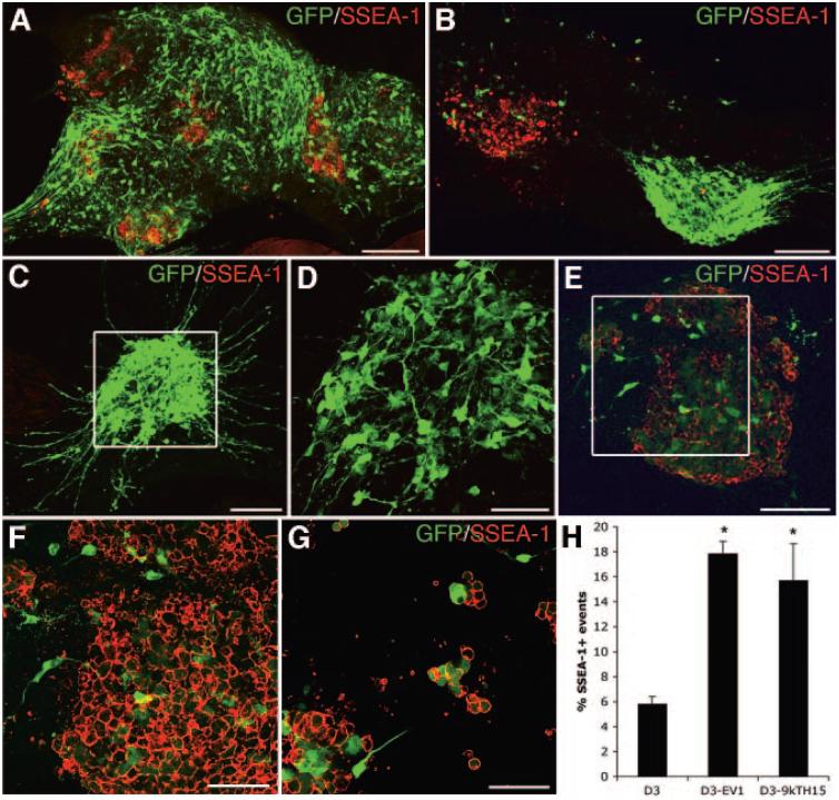
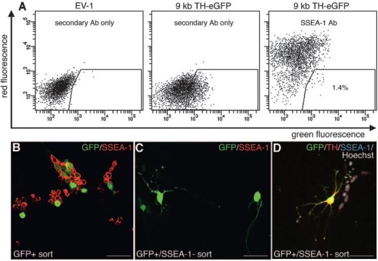
Transplantation of mouse embryonic stem (mES) cells can restore function in Parkinson disease models, but can generate teratomas. Purification of dopamine neurons derived from embryonic stem cells by fluorescence-activated cell sorting (FACS) could provide a functional cell population for transplantation while eliminating the risk of teratoma formation. Here we used the tyrosine hydroxylase (TH) promoter to drive enhanced green fluorescent protein (eGFP) expression in mES cells. First, we evaluated 2.5-kilobase (kb) and 9-kb TH promoter fragments and showed that clones generated using the 9-kb fragment produced significantly more eGFP+/TH+ neurons. We selected the 9-kb TH clone with the highest eGFP/TH overlap for further differentiation, FACS, and transplantation experiments. Grafts contained large numbers of eGFP+ dopamine neurons of an appropriate phenotype. However, there were also numerous eGFP+ cells that did not express TH and did not have a neuronal morphology. In addition, we found cells in the grafts representing all three germ layers. Based on these findings, we examined the expression of stem cell markers in our eGFP+ population. We found that a majority of eGFP+ cells were stage-specific embryonic antigen-positive (SSEA-1+) and that the genetically engineered clones contained more SSEA-1+ cells after differentiation than the original D3 mES cells. By negative selection of SSEA-1, we could isolate a neuronal eGFP+ population of high purity. These results illustrate the complexity of using genetic selection to purify mES cell-derived dopamine neurons and provide a comprehensive analysis of cell selection strategies based on tyrosine hydroxylase expression. Disclosure of potential conflicts of interest is found at the end of this article.


Figures

Figure 1
Figure 1
Evaluation of TH-eGFP promoter constructs in mouse embryonic stem (mES) cells during in vitro differentiation. (A, B): Overlap in eGFP and TH expression in a (A) 2.5-kilobase (kb) TH-GFP clone and in a (B) 9-kb TH-eGFP clone after 14 days of differentiation on PA6. Cultures of 9-kb TH-eGFP clones contained many more eGFP+/TH+ cells compared with the 2.5-kb TH-GFP cultures (yellow co-expression). (C): FACS analysis for eGFP+ events within an FSC/SSC gate of naïve D3 mES cells autofluorescence), one 2.5-kb TH-eGFP clone (number 91) and three 9-kb TH-eGFP clones (numbers 15, 41, and 93) after 9 days of differentiation on PA6. All 9-kb TH clones showed an expected higher proportion of eGFP+ events compared with the naïve D3 mES cell line (*, p < .01 to p < .001; analysis of variance [ANOVA]) and to the 2.5-kb clone (#, p < .01 to p < .001; ANOVA). The 2.5-kb TH-eGFP clone was not significantly different from the D3 cells (*, p < .05 to p < .001; ANOVA). Scale bars = 50 μm (A, B). Abbreviations: eGFP, enhanced green fluorescent protein; FSC, forward scatter; GFP, green fluorescent protein; k, kilobase; SSC, side scatter; TH, tyrosine hydroxylase.
Figure 2
Figure 2
Comparison of MS5- and PA6-based in vitro differentiation protocols. (A-H): The 9-kilobase (kb) TH-eGFP mouse embryonic stem cells were analyzed in vitro after 9 days of differentiation using either the PA6 or MS5-based protocols. (A), enlarged in (B): eGFP and TuJ1 overlap was high using either protocol, although it was significantly higher using the MS5-based protocol (PA6, 94.9% ± 0.2% [SEM]; MS5, 98.9% ± 0.7% [SEM]) (yellow co-expression) (G). eGFP and TH overlap was significantly higher using the MS5-based compared with the PA6-based protocol (C, D, G) (PA6, 25.7% ± 6.8% [SEM]; MS5, 60.9% ± 1.6% [SEM]) (yellow co-expression). Pax2 immunofluorescent staining (E) showed the mid-hindbrain characteristic of the MS5 culture, with very little overlap between eGFP and Pax2, as anticipated based on the different temporal expression of TH and Pax2 during normal development. (F): There were few DBH+ cells generated in either PA6- or MS5-based protocols, and the DBH staining did not appear to overlap with eGFP expression. Bar graphs depicting overlap between GFP and TuJ1 expression (G) and eGFP and TH expression (H) in cells differentiated using PA6-based (white bars) or MS5-based (black bars) protocol for 9 days (*, p < .01, unpaired t test). Scale bars = 100 μm (A) and 50 μm (B-F). Abbreviations: DBH, dopamine β-hydroxylase; eGFP, enhanced green fluorescent protein; GFP, green fluorescent protein; Pax2, paired box gene 2; TH, tyrosine hydroxylase; TuJ1, class III β-tubulin.
Figure 3
Figure 3
Short-term transplantation and in vivo analysis of eGFP-sorted cells. (A-O): naïve mice were transplanted with cells sorted for eGFP expression. Grafts were analyzed 4 weeks post-transplantation. (A): Low-power microphotograph of a graft, showing large numbers of TH+ neurons (the nickel-enhanced 3,3′-diaminobenzidine products appear grayish black; the boxed area is shown enlarged in [B]). Most TH+ neurons appeared to co-express eGFP (C) (co-expression appears yellow; the boxed area is enlarged in [D]); however, some cells expressed only eGFP or only TH. A large proportion of the eGFP+/TH cells also showed overlapping expression with AADC (arrowheads) (co-expression appears purple) (E-H). Many eGFP+/TH+ cells showed overlapping GIRK2 immunoreactivity (arrowheads) (co-expression appears purple), but not all (I-L). Pitx3 was expressed in the nucleus of most eGFP+ cells (M-O). Scale bars = 150 μm (A), 30 μm (B), 100 μm (C), and 50 μm (D, H, L, O) (scale bar in [H] applies to [E-G], [L] applies to [I-K], and [O] applies to [M, N]). Abbreviations: AADC, aromatic L-amino acid decarboxylase; GFP, green fluorescent protein; GIRK2, G protein-activated inwardly rectifying potassium channel 2; Pitx3, paired-like homeodomain transcription factor 3; TH, tyrosine hydroxylase.
Figure 4
Figure 4
Long-term xenotransplantation and in vivo analysis of eGFP-sorted cells. (A-N): eGFP+ cell transplants into 6-hydroxydopamine-lesioned rats were analyzed 10 weeks post-transplantation. (A): Low-power microphotograph of a graft, showing large numbers of TH+ neurons (the nickel-enhanced DAB products appear grayish black; the boxed area to the left is shown enlarged in [B], and the boxed area to the right is shown enlarged in [C]). Some of the grafted TH neurons extend neurites into the host striatum (C). (D): Many TH+ neurons were eGFP+ (co-expression appears yellow). (E): Bar graph depicting the percentage of total TH+ cells that were eGFP+ (white bar; 60.7% ± 6.4% [SEM]) or eGFP- (black bar; 39.3% ± 6.3% [SEM]). The two groups were not significantly different (p > .05, paired t test) due to the high variability in overlap between TH and eGFP between different grafts. Many of the eGFP+ neurons displayed characteristics of midbrain dopamine neurons, as shown by the overlapping expression with AADC (F-H) and Pitx3 (I-K). Grafts contained all three germ layers, as visualized by staining for cytokeratin (L), myosin (M), and villin (N), but no overlapping expression with eGFP was detected. Scale bars = 200 μm (A), 50 μm (B, C, F-N), and 25 μm (D). Abbreviations: AADC, aromatic L-amino acid decarboxylase; eGFP, enhanced green fluorescent protein; GFP, green fluorescent protein; Pitx3, paired-like homeodomain transcription factor 3; TH, tyrosine hydroxylase.
Figure 5
Figure 5
Identification of proliferative eGFP+ cells in the grafts. (A-L): Analysis of mouse grafts 4 weeks post-transplantation showed that there were cluster of SSEA-1+/eGFP+ cells in some grafts ([A]; the boxed area is shown enlarged in [B-D]). Further analysis of the rat grafts 10 weeks post-transplantation showed that many eGFP+ cells were Ki67+ as well as SSEA-1+ ([E-I]; [F-H] show enlargements of [E], and [I] is an orthogonal view; co-expression of Ki67 and eGFP appears yellow). Staining for neural progenitors showed that the grafts contained many nestin+ cells (K, L) but that few of these cells were eGFP+ (J, L). Scale bars = 100 μm (A, E) and 50 μm (D, H, L) (scale bar in [D] applies to [B, C], that in [H] applies to [F, G], and that in [L] applies to [J, K]). Abbreviations: GFP, green fluorescent protein; SSEA, stage-specific embryonic antigen.
Figure 6
Figure 6
In vitro analysis of SSEA-1 expression in differentiated mouse embryonic stem (mES) cell cultures. (A-H): Analysis of in vitro-differentiated 9-kilobase (kb) TH-eGFP cells showed that most colonies contained many eGFP+ neurons and few SSEA-1+ cells and no or very few cells that were eGFP+/SSEA-1+ ([A-E]; [D] is an enlargement of [C]). Some colonies, however, contained a majority of SSEA-1+ cells ([B, E, F]; [F] is an enlargement of [E]), and in some instances, these cells were SSEA-1+/eGFP+ (E, F). The eGFP+/SSEA-1+ cells appeared to have a lower expression of eGFP than the eGFP+ cells with neuronal morphology before dissociation (C, F). The expression of SSEA-1 did not appear to be correlated with the size of the colony (B). Dissociation of cultures followed by replating showed that most cell surviving this procedure were SSEA-1+ (G). Fluorescence-activated cell sorting analysis showed that 9-kb TH-eGFP cells, as well as the EV1-transfected mES cells, contained a higher number of SSEA-1+ cells than the original D3 mES cells used for the genetic manipulations (H). Scale bars = 100 μm (A-C, E) and 50 μm (D, F, G). Abbreviations: k, kilobase; EV1, empty vector; GFP, green fluorescent protein; SSEA, stage-specific embryonic antigen; TH, tyrosine hydroxylase.
Figure 7
Figure 7
Purification of neurons from in vitro-differentiated TH-eGFP cultures by negative selection for SSEA-1. Live stain and fluorescence-activated cell sorting (FACS) for eGFP+ and SSEA-1, after in vitro differentiation on MS5 for 9 days, showed that most eGFP+ cells were SSEA1+, although a small population of cells that were eGFP+/SSEA-1- could be isolated (A). The EV-1-transfected mouse embryonic stem cell clone, differentiated for the same length of time as the 9-kb TH-eGFP clone, was used as a negative control for fluorescence (A). Plating of cells sorted only for eGFP fluorescence resulted in a population consisting mainly of SSEA-1+ cells of a non-neuronal morphology (2 days in vitro post-FACS) (B). Cells negatively sorted for SSEA-1 and positively for eGFP gave a population of high neuronal purity (A, C, D). Some of the eGFP+ neurons from this double sort also expressed TH (2 days in vitro post-FACS) (D). Abbreviations: Ab, antibody; eGFP, enhanced green fluorescent protein; EV-1, empty vector; GFP, green fluorescent protein; kb, kilobase; SSEA, stage-specific embryonic antigen; TH, tyrosine hydroxylase.

References

    1. Bjorklund LM, Sanchez-Pernaute R, Chung S, et al. Embryonic stem cells develop into functional dopaminergic neurons after transplantation in a Parkinson rat model. Proc Natl Acad Sci U S A. 2002;99:2344–2349. - PMC - PubMed
    1. Kim JH, Auerbach JM, Rodriguez-Gomez JA, et al. Dopamine neurons derived from embryonic stem cells function in an animal model of Parkinson's disease. Nature. 2002;418:50–56. - PubMed
    1. Barberi T, Klivenyi P, Calingasan NY, et al. Neural subtype specification of fertilization and nuclear transfer embryonic stem cells and application in parkinsonian mice. Nat Biotechnol. 2003;21:1200–1207. - PubMed
    1. Ben-Hur T, Idelson M, Khaner H, et al. Transplantation of human embryonic stem cell-derived neural progenitors improves behavioral deficit in Parkinsonian rats. STEM CELLS. 2004;22:1246–1255. - PubMed
    1. Arnhold S, Klein H, Semkova I, et al. Neurally selected embryonic stem cells induce tumor formation after long-term survival following engraftment into the subretinal space. Invest Ophthalmol Vis Sci. 2004;45:4251–4255. - PubMed

Publication types