Comparative Study
. 2007 Feb;8(2):158-64.
doi: 10.1038/sj.embor.7400890. Epub 2007 Jan 19.

PATJ regulates directional migration of mammalian epithelial cells

Affiliations
Comparative Study

PATJ regulates directional migration of mammalian epithelial cells

Kunyoo Shin et al. EMBO Rep. 2007 Feb.

Abstract

Directional migration is important in wound healing by epithelial cells. Recent studies have shown that polarity proteins such as mammalian Partitioning-defective 6 (Par6), atypical protein kinase C (aPKC) and mammalian Discs large 1 (Dlg1) are crucial not only for epithelial apico-basal polarity, but also for directional movement. Here, we show that the protein associated with Lin seven 1 (PALS1)-associated tight junction protein (PATJ), another evolutionarily conserved polarity protein, is also required for directional migration by using a wound-induced migration assay. In addition, we found that aPKC and Par3 localize to the leading edge during migration of epithelia and that PATJ regulates their localization. Furthermore, our results show that microtubule-organizing centre orientation is disrupted in PATJ RNA interference (RNAi) MDCKII (Madin-Darby canine kidney II) cells during migration. Together, our data indicate that PATJ controls directional migration by regulating the localization of aPKC and Par3 to the leading edge. The migration defect in PATJ RNAi cells seems to be due to the disorganization of the microtubule network induced by mislocalization of polarity proteins.


Figures
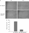
Figure 1
Figure 1
PATJ is required for the migration of epithelial cells. (A) Wound-closure migration assays were performed with wild-type (WT) and PATJ RNAi, MDCKII cells. Wound areas are outlined. Scale bar, 100 μm. (B) Quantification of epithelial migration after wounding. Wild-type MDCKII cells show 87.4±1.4% wound closure, whereas PATJ RNAi MDCKII cells show 15.4±3.2% wound closure 17 h after wounding; P<0.0001, unpaired t-test. Relative areas of the wound were measured in three independent experiments. Standard deviations are shown as error bars (n=3). MDCKII, Madin–Darby canine kidney II; PALS1, protein associated with Lin seven 1; Par3, partitioning-defective 3; PATJ, PALS1-associated tight junction protein; RNAi, RNA interference.
Figure 2
Figure 2
PATJ is required for the localization of aPKC and Par3 to the leading edge of migrating epithelial cells. PATJ, aPKC and Par3 were immunostained 6 h after wounding in wild-type (WT; A), PATJ RNAi (B) and EGFP–PATJ full-length (FL) rescue PATJ RNAi MDCKII cells (C). Arrows show the localization of PATJ, aPKC and Par3 to the leading edge. Nuclei are shown in blue. Scale bar, 10 μm. (D) Quantification of aPKC and Par3 localization during migration. The score of Par3 localization decreases from 101.3±3.5 (WT MDCKII) to 53.5±6.7 (PATJ RNAi MDCKII cells); P<0.0005, unpaired t-test. The score of aPKC localization decreases from 75.3±5.7 (WT MDCKII) to 26.0±4.6 (PATJ RNAi MDCKII cells); P<0.0005, unpaired t-test. Standard deviations are shown as error bars (n=3). aPKC, atypical protein kinase C; EGFP, enhanced green fluorescent protein; MDCKII, Madin–Darby canine kidney II; PALS1, protein associated with Lin seven 1; Par3, Partitioning-defective 3; PATJ, PALS1-associated tight junction protein; RNAi, RNA interference.
Figure 3
Figure 3
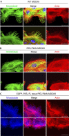
Organization of microtubules is disrupted in PATJ RNAi MDCKII cells during wound healing. (A,B) Microtubules (green) and actin (red) were visualized in wild-type (WT; A) and PATJ RNAi (B) MDCKII cells 6 h after wounding. Merged images are shown in the middle column. Scale bars, 10 μm. (C) Microtubules (blue) and actin (red) were visualized in EGFP–PATJ full-length (FL) rescue PATJ RNAi MDCKII cells. Scale bars, 10 μm. EGFP, enhanced green fluorescent protein; MDCKII, Madin–Darby canine kidney II; PALS1, protein associated with Lin seven 1; PATJ, PALS1-associated tight junction protein; RNAi, RNA interference.
Figure 4
Figure 4
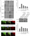
Structure–function analysis of PATJ. (A) Wound closure migration assays were performed with EGFP, EGFP–PATJ full length (FL), EGFP-PATJ ΔL27, EGFP–PATJ ΔPDZ1–5 and EGFP–PATJ ΔPDZ6–10 rescue PATJ RNAi MDCKII cells. Wound areas are outlined. Scale bar, 100 μm. (B) Quantification of epithelial migration with mutant PATJ rescue cell lines after wounding. Relative areas of the wound were measured in three independent experiments. Standard deviations are shown as error bars (n=3). Results as percentage wound closure at 17 h are as follows: EGFP rescue PATJ RNAi: 11.2±4.1%; EGFP PATJ FL rescue PATJ RNAi: 56.1±3.6%; EGFP PATJ ΔL27 rescue PATJ RNAi: 33.9±3.8%; EGFP PATJ ΔPDZ1–5 rescue PATJ RNAi: 24.9±3.8%; EGFP PATJ ΔPDZ6–10 rescue PATJ RNAi: 19.3±1.2%. (C) Immunoprecipitation of EGFP–PATJ mutant proteins followed by western blot for Par3. (D) Par3 was immunostained 6 h after wounding with EGFP–PATJ mutant rescue PATJ RNAi cell lines. Scale bar, 10 μm. (E) Quantification of Par3 localization during migration of EGFP–PATJ mutant rescue PATJ RNAi MDCKII cells. EGFP, enhanced green fluorescent protein; MDCKII, Madin–Darby canine kidney II; PALS1, protein associated with Lin seven 1; Par3, Partitioning-defective 3; PATJ, PALS1-associated tight junction protein; RNAi, RNA interference.

References

    1. Bazzoni G, Martinez-Estrada OM, Orsenigo F, Cordenonsi M, Citi S, Dejana E (2000) Interaction of junctional adhesion molecule with the tight junction components ZO-1, cingulin, and occludin. J Biol Chem 275: 20520–20526 - PubMed
    1. Berrueta L, Tirnauer JS, Schuyler SC, Pellman D, Bierer BE (1999) The APC-associated protein EB1 associates with components of the dynactin complex and cytoplasmic dynein intermediate chain. Curr Biol 9: 425–428 - PubMed
    1. Chen X, Macara IG (2005) Par-3 controls tight junction assembly through the Rac exchange factor Tiam1. Nat Cell Biol 7: 262–269 - PubMed
    1. Craig AM, Banker G (1994) Neuronal polarity. Annu Rev Neurosci 17: 267–310 - PubMed
    1. Ebnet K, Schulz CU, Meyer Zu Brickwedde MK, Pendl GG, Vestweber D (2000) Junctional adhesion molecule interacts with the PDZ domain-containing proteins AF-6 and ZO-1. J Biol Chem 275: 27979–27988 - PubMed

Publication types