

Inheritance of mitochondrial DNA recombinants in double-heteroplasmic families: potential implications for phylogenetic analysis
- PMID: 17236134
- PMCID: PMC1785346
- DOI: 10.1086/511282
Inheritance of mitochondrial DNA recombinants in double-heteroplasmic families: potential implications for phylogenetic analysis
Abstract
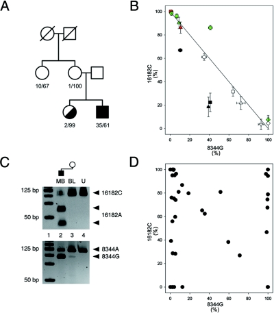
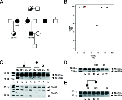
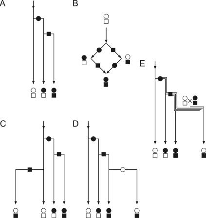
Recently, somatic recombination of human mitochondrial DNA (mtDNA) was discovered in skeletal muscle. To determine whether recombinant mtDNA molecules can be transmitted through the germ line, we investigated two families, each harboring two inherited heteroplasmic mtDNA mutations. Using allele-specific polymerase chain reaction and single-cell and single-molecule mutational analyses, we discovered, in both families, all four possible allelic combinations of the two heteroplasmic mutations (tetraplasmy), the hallmark of mtDNA recombination. We strongly suggest that these recombinant mtDNA molecules were inherited rather than de novo generated somatically, because they (1) are highly abundant and (2) are present in different tissues of maternally related family members, including young individuals. Moreover, the comparison of the complete mtDNA sequence of one of the families with database sequences revealed an irregular, nontreelike pattern of mutations, reminiscent of a reticulation. We therefore propose that certain reticulations of the human mtDNA phylogenetic tree might be explained by recombination of coexisting mtDNA molecules harboring multiple mutations.
Figures
Similar articles
-
A MERRF/MELAS overlap syndrome associated with a new point mutation in the mitochondrial DNA tRNA(Lys) gene.Eur J Hum Genet. 1993;1(1):80-7. doi: 10.1159/000472390. Eur J Hum Genet. 1993. PMID: 8069654
-
The A to G transition at nt 3243 of the mitochondrial tRNALeu(UUR) may cause an MERRF syndrome.J Neurol Neurosurg Psychiatry. 1996 Jul;61(1):47-51. doi: 10.1136/jnnp.61.1.47. J Neurol Neurosurg Psychiatry. 1996. PMID: 8676159 Free PMC article.
-
Analysis of the tissue distribution and inheritance of heteroplasmic mitochondrial DNA point mutation by denaturing gradient gel electrophoresis in MERRF syndrome.Neuromuscul Disord. 1992;2(5-6):323-30. doi: 10.1016/s0960-8966(06)80003-9. Neuromuscul Disord. 1992. PMID: 1300181
-
[The correlation of the heteroplasmy of mtDNA and clinicopathological findings in the patients with mitochondrial encephalomyopathies].Nihon Rinsho. 1997 Dec;55(12):3270-6. Nihon Rinsho. 1997. PMID: 9436449 Review. Japanese.
-
[Mitochondrial DNA mutations and three major forms of mitochondrial myopathies: CPEO, MELAS and MERRF].Nihon Rinsho. 1997 Dec;55(12):3259-64. Nihon Rinsho. 1997. PMID: 9436447 Review. Japanese.
Cited by
-
How good are indirect tests at detecting recombination in human mtDNA?G3 (Bethesda). 2013 Jul 8;3(7):1095-104. doi: 10.1534/g3.113.006510. G3 (Bethesda). 2013. PMID: 23665874 Free PMC article.
-
Distinct patterns of mitochondrial genome diversity in bonobos (Pan paniscus) and humans.BMC Evol Biol. 2010 Sep 2;10:270. doi: 10.1186/1471-2148-10-270. BMC Evol Biol. 2010. PMID: 20813043 Free PMC article.
-
No recombination of mtDNA after heteroplasmy for 50 generations in the mouse maternal germline.Nucleic Acids Res. 2014 Jan;42(2):1111-6. doi: 10.1093/nar/gkt969. Epub 2013 Oct 25. Nucleic Acids Res. 2014. PMID: 24163253 Free PMC article.
-
A wide range of 3243A>G/tRNALeu(UUR) (MELAS) mutation loads may segregate in offspring through the female germline bottleneck.PLoS One. 2014 May 7;9(5):e96663. doi: 10.1371/journal.pone.0096663. eCollection 2014. PLoS One. 2014. PMID: 24805791 Free PMC article.
-
Linear DNA-driven recombination in mammalian mitochondria.Nucleic Acids Res. 2024 Apr 12;52(6):3088-3105. doi: 10.1093/nar/gkae040. Nucleic Acids Res. 2024. PMID: 38300793 Free PMC article.
References
Web Resources
-
- GenBank, http://www.ncbi.nlm.nih.gov/Genbank/ (for the complete mitochondrial sequences of the patient with the A3243G/G16428A mutations [accession number DQ862536] and of the sister of the patient with the A8344G/A16182C mutations [accession number DQ862537] and Homo sapiens isolates F128 [accession numbers AY339529] and F71 [accession number AY339472])
-
- ImageJ, http://rsb.info.nih.gov/ij/ (for Image Analysis Software)
-
- mtDB–Human Mitochondrial Genome Database, http://www.genpat.uu.se/mtDB/
-
- Online Mendelian Inheritance in Man (OMIM), http://www.ncbi.nlm.nih.gov/Omim/ (for MERRF syndrome and MELAS syndrome)
References
-
- Rokas A, Ladoukakis E, Zouros E (2003) Animal mitochondrial DNA recombination revisited. Trends Ecol Evol 18:411–41710.1016/S0169-5347(03)00125-3 - DOI
Publication types
MeSH terms
Substances
Associated data
- Actions
- Actions
- Actions
- Actions
Grants and funding
LinkOut - more resources
Full Text Sources
