

Mutations in HOXD13 underlie syndactyly type V and a novel brachydactyly-syndactyly syndrome
- PMID: 17236141
- PMCID: PMC1785357
- DOI: 10.1086/511387
Mutations in HOXD13 underlie syndactyly type V and a novel brachydactyly-syndactyly syndrome
Abstract
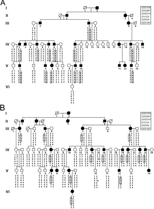
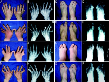
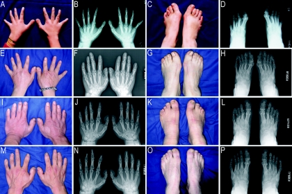
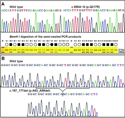
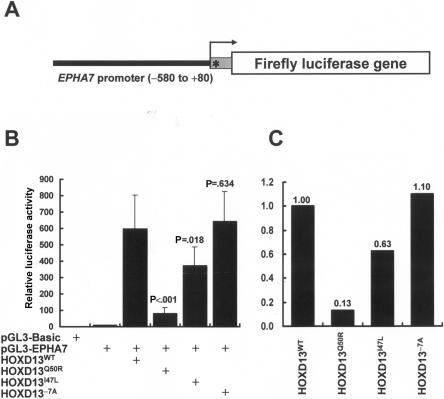
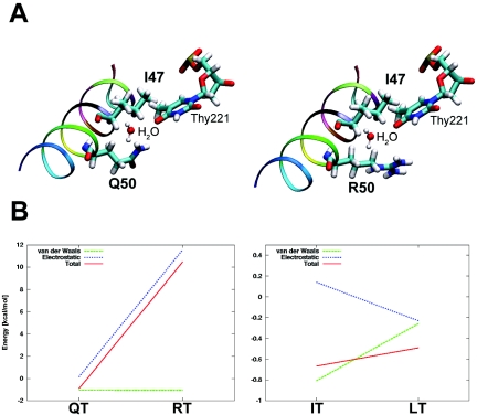
HOXD13, the homeobox-containing gene located at the most 5' end of the HOXD cluster, plays a critical role in limb development. It has been shown that mutations in human HOXD13 can give rise to limb malformations, with variable expressivity and a wide spectrum of clinical manifestations. Polyalanine expansions in HOXD13 cause synpolydactyly, whereas amino acid substitutions in the homeodomain are associated with brachydactyly types D and E. We describe two large Han Chinese families with different limb malformations, one with syndactyly type V and the other with limb features overlapping brachydactyly types A4, D, and E and mild syndactyly of toes 2 and 3. Two-point linkage analysis showed LOD scores >3 (theta =0) for markers within and/or flanking the HOXD13 locus in both families. In the family with syndactyly type V, we identified a missense mutation in the HOXD13 homeodomain, c.950A-->G (p.Q317R), which leads to substitution of the highly conserved glutamine that is important for DNA-binding specificity and affinity. In the family with complex brachydactyly and syndactyly, we detected a deletion of 21 bp in the imperfect GCN (where N denotes A, C, G, or T) triplet-containing exon 1 of HOXD13, which results in a polyalanine contraction of seven residues. Moreover, we found that the mutant HOXD13 with the p.Q317R substitution was unable to transactivate the human EPHA7 promoter. Molecular modeling data supported these experimental results. The calculated interactions energies were in agreement with the measured changes of the activity. Our data established the link between HOXD13 and two additional limb phenotypes--syndactyly type V and brachydactyly type A4--and demonstrated that a polyalanine contraction in HOXD13, most likely, led to other digital anomalies but not to synpolydactyly. We suggest the term "HOXD13 limb morphopathies" for the spectrum of limb disorders caused by HOXD13 mutations.
Figures
References
Web Resources
-
- GenBank, http://www.ncbi.nlm.nih.gov/Genbank/ (for HOXD13 [accession number NM_000523], HOXD9 [accession number NM_014213], HOXD10 [accession number NM_002148], HOXD11 [accession number NM_021192], HOXD12 [accession number NM_021193], and EPHA7 [accession number NM_004440])
-
- Homeodomain Resource, http://genome.nih.gov/homeodomain/
-
- Human Genome Browser Gateway, http://genome.ucsc.edu/cgi-bin/hgGateway
-
- Online Mendelian Inheritance in Man (OMIM), http://www.ncbi.nlm.nih.gov/Omim/ (for HOXD13, SPD, BDD, BDE, syndactyly type V, BDA4, syndactyly type I, Guttmacher syndrome, and hand-foot-genital syndrome)
-
- Protein Data Bank, http://www.rcsb.org/pdb/ (for the Antp [ID 9ANT] homeodomain DNA structure)
References
-
- Capecchi MR (1997) Hox genes and mammalian development. Cold Spring Harb Symp Quant Biol 62:273–281 - PubMed
Publication types
MeSH terms
Substances
Associated data
- Actions
- Actions
- Actions
- Actions
- Actions
- Actions
- Actions
Grants and funding
LinkOut - more resources
Full Text Sources
Other Literature Sources
Molecular Biology Databases
Research Materials
Miscellaneous
