. 2007 Feb;80(2):372-7.
doi: 10.1086/511527. Epub 2006 Dec 28.

The reduced expression of the HADH2 protein causes X-linked mental retardation, choreoathetosis, and abnormal behavior

Affiliations

The reduced expression of the HADH2 protein causes X-linked mental retardation, choreoathetosis, and abnormal behavior

Claus Lenski et al. Am J Hum Genet. 2007 Feb.

Abstract

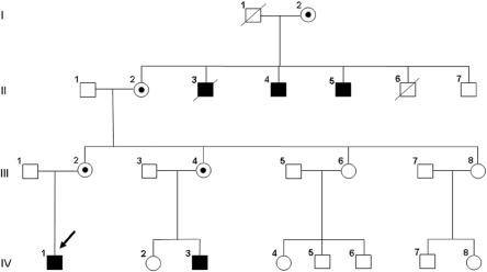
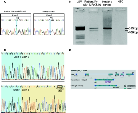
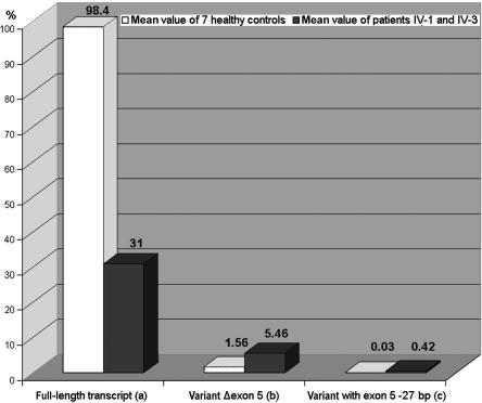
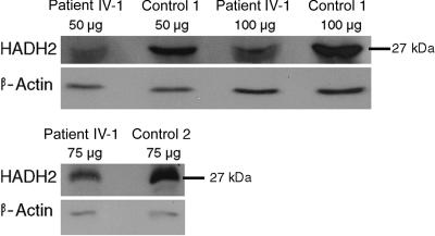
Recently, we defined a new syndromic form of X-linked mental retardation in a 4-generation family with a unique clinical phenotype characterized by mild mental retardation, choreoathetosis, and abnormal behavior (MRXS10). Linkage analysis in this family revealed a candidate region of 13.4 Mb between markers DXS1201 and DXS991 on Xp11; therefore, mutation analysis was performed by direct sequencing in most of the 135 annotated genes located in the region. The gene (HADH2) encoding L-3-hydroxyacyl-CoA dehydrogenase II displayed a sequence alteration (c.574 C-->A; p.R192R) in all patients and carrier females that was absent in unaffected male family members and could not be found in 2,500 control X chromosomes, including in those of 500 healthy males. The silent C-->A substitution is located in exon 5 and was shown by western blot to reduce the amount of HADH2 protein by 60%-70% in the patient. Quantitative in vivo and in vitro expression studies revealed a ratio of splicing transcript amounts different from those normally seen in controls. Apparently, the reduced expression of the wild-type fragment, which results in the decreased protein expression, rather than the increased amount of aberrant splicing fragments of the HADH2 gene, is pathogenic. Our data therefore strongly suggest that reduced expression of the HADH2 protein causes MRXS10, a phenotype different from that caused by 2-methyl-3-hydroxybutyryl-CoA dehydrogenase deficiency, which is a neurodegenerative disorder caused by missense mutations in this multifunctional protein.


Figures

Figure  1.
Figure 1.
Pedigree of the MRXS10-affected family. Affected males are indicated by black boxes, the obligate carrier females by circles with dots. The index patient (IV-1) is marked with an arrow.
Figure  2.
Figure 2.
A, Silent c.574C→A substitution in exon 5 of HADH2. Left, Sequence of MRXS10-affected patient IV-1, who harbors the c.574C→A substitution. Right, Wild-type sequence of a healthy control. B, RT-PCR amplification of the HADH2 gene through use of primers HADH2-cF2 and HADH2-cR2. In patient IV-1 (lane 2), the amplification results in a 515-bp wild-type fragment and in a second, shorter fragment of 406 bp. In the healthy control (lane 3), only the wild-type 515-bp fragment is visible. The negative template control is shown in lane 4. Molecular weight marker LSV is shown in lane 1. C, cDNA sequences of the exon boundaries (between exons 4 and 5) of the 515-bp fragment with exon 5 (top) and of the exon boundaries (exons 4–6) of the 416-bp fragment lacking exon 5 (bottom). D, Genomic structure of HADH2. The gene consists of six coding exons, which result in a 261-aa protein with a conserved short-chain dehydrogenase domain at amino acid positions 28–257 (hatched boxes). The c.574C→A substitution is located in exon 5. In the transcript variant lacking exon 5, a premature stop codon is created in exon 6. The position of the primers used for cDNA amplification, as shown in panel B, are indicated by “cF2” and “cR2.'
Figure  3.
Figure 3.
Transcript quantification applying real-time PCR. Whereas the full-length transcript is reduced to one-third in patients IV-1 and IV-3, as compared with the mean value of seven healthy controls (a), the Δexon 5 variant is increased 3.5-fold in the patient (b), and the transcript variant that lacks 27 bp of the 3′ end of exon 5 is increased 14-fold in the patients (c). RNA samples from the two patients and the seven controls were reverse transcribed (cDNA Synthesis Kit [Amersham]).
Figure  4.
Figure 4.
Detection of the HADH2 protein by western-blot analysis. Amounts between 50 and 100 μg of total protein were loaded. As reference, protein β-actin was detected. The ∼27-kDa bands representing the HADH2 protein of patient IV-1 with MRXS10 show a weaker signal than do the bands of control individuals 1 and 2.

References

Web Resources

    1. GenBank, http://www.ncbi.nlm.nih.gov/Genbank/ (for HADH2 [accession number NM_004493])
    1. Online Mendelian Inheritance in Man (OMIM), http://www.ncbi.nlm.nih.gov/Omim/ (for MRXS10, HADH2, FTDP-17, XMRE, MHBD deficiency, PD, and AD)

References

    1. Chelly J, Mandel JL (2001) Monogenic causes of X-linked mental retardation. Nat Rev Genet 2:669–68010.1038/35088558 - DOI - PubMed
    1. Stevenson RE, Schwartz, CE (2002) Clinical and molecular contributions to the understanding of X-linked mental retardation. Cytogenet Genome Res 99:265–27510.1159/000071603 - DOI - PubMed
    1. Ropers HH, Hamel BC (2005) X-linked mental retardation. Nat Rev Genet 6:46–5710.1038/nrg1501 - DOI - PubMed
    1. Stevenson RE (2005) Advances in X-linked mental retardation. Curr Opin Pediatr 17:720–72410.1097/01.mop.0000184290.57525.fb - DOI - PubMed
    1. Reyniers E, Van Bogaert NP, Vits L, Pauly F, Fransen E, Van Regemorter V, Kooy FR (1999) A new neurological syndrome with mental retardation, choreoathetosis, and abnormal behavior maps to chromosome Xp11. Am J Hum Genet 65:1406–1412 - PMC - PubMed

Publication types

Substances

Associated data