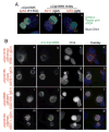
Localization of the hypothetical protein Cpn0585 in the inclusion membrane of Chlamydia pneumoniae-infected cells
- PMID: 17236746
- PMCID: PMC1850435
- DOI: 10.1016/j.micpath.2006.11.006
Localization of the hypothetical protein Cpn0585 in the inclusion membrane of Chlamydia pneumoniae-infected cells
Abstract
Cpn0585, encoded by a hypothetical open reading frame in Chlamydia pneumoniae genome, was detected in the inclusion membrane during C. pneumoniae infection using both polyclonal and monoclonal antibodies raised with Cpn0585 fusion protein. The anti-Cpn0585 antibodies specifically recognized the endogenous Cpn0585 without cross-reacting with IncA (a known inclusion membrane protein of C. pneumoniae) or other control antigens. A homologue of Cpn0585 in the C. caviae species (encoded by the ORF CCA00156) was also localized in the inclusion membrane of the C. caviae-infected cells. The Cpn0585 protein became detectable 24h while CCA00156 as early as 8h after infection. Once expressed, both proteins remained in the inclusion membrane throughout the rest of infection course.
Figures


Similar articles
-
Chlamydia pneumoniae inclusion membrane protein Cpn0585 interacts with multiple Rab GTPases.Infect Immun. 2007 Dec;75(12):5586-96. doi: 10.1128/IAI.01020-07. Epub 2007 Oct 1. Infect Immun. 2007. PMID: 17908815 Free PMC article.
-
Hypothetical protein Cpn0308 is localized in the Chlamydia pneumoniae inclusion membrane.Infect Immun. 2007 Jan;75(1):497-503. doi: 10.1128/IAI.00935-06. Epub 2006 Nov 13. Infect Immun. 2007. PMID: 17101661 Free PMC article.
-
[Effector proteins of Clamidia].Mol Biol (Mosk). 2009 Nov-Dec;43(6):963-83. Mol Biol (Mosk). 2009. PMID: 20088373 Review. Russian.
-
A secondary structure motif predictive of protein localization to the chlamydial inclusion membrane.Cell Microbiol. 2000 Feb;2(1):35-47. doi: 10.1046/j.1462-5822.2000.00029.x. Cell Microbiol. 2000. PMID: 11207561
-
Chlamydia pneumoniae: modern insights into an ancient pathogen.Trends Microbiol. 2013 Mar;21(3):120-8. doi: 10.1016/j.tim.2012.10.009. Epub 2012 Dec 5. Trends Microbiol. 2013. PMID: 23218799 Review.
Cited by
-
Multi-genome identification and characterization of chlamydiae-specific type III secretion substrates: the Inc proteins.BMC Genomics. 2011 Feb 16;12:109. doi: 10.1186/1471-2164-12-109. BMC Genomics. 2011. PMID: 21324157 Free PMC article.
-
Molecular mechanisms of host-pathogen interactions and their potential for the discovery of new drug targets.Curr Drug Targets. 2008 Feb;9(2):150-7. doi: 10.2174/138945008783502449. Curr Drug Targets. 2008. PMID: 18288966 Free PMC article. Review.
-
Recombinant Cpn 0810 stimulates proinflammatory cytokine expression and apoptosis in human monocytes.Exp Ther Med. 2015 Feb;9(2):459-463. doi: 10.3892/etm.2014.2111. Epub 2014 Dec 5. Exp Ther Med. 2015. PMID: 25574216 Free PMC article.
-
The Chlamydia pneumoniae Inclusion Membrane Protein Cpn1027 Interacts with Host Cell Wnt Signaling Pathway Regulator Cytoplasmic Activation/Proliferation-Associated Protein 2 (Caprin2).PLoS One. 2015 May 21;10(5):e0127909. doi: 10.1371/journal.pone.0127909. eCollection 2015. PLoS One. 2015. PMID: 25996495 Free PMC article.
-
Hypothetical protein Cpn0423 triggers NOD2 activation and contributes to Chlamydia pneumoniae-mediated inflammation.BMC Microbiol. 2017 Jul 11;17(1):153. doi: 10.1186/s12866-017-1062-y. BMC Microbiol. 2017. PMID: 28693414 Free PMC article.
References
-
- Stephens RS, Kalman S, Lammel C, et al. Genome sequence of an obligate intracellular pathogen of humans: Chlamydia trachomatis. Science. 1998;282:754–9. - PubMed
-
- Everett KD, Bush RM, Andersen AA. Emended description of the order Chlamydiales, proposal of Parachlamydiaceae fam. nov. and Simkaniaceae fam. nov., each containing one monotypic genus, revised taxonomy of the family Chlamydiaceae, including a new genus and five new species, and standards for the identification of organisms. Int J Syst Bacteriol. 1999;49(Pt 2):415–40. - PubMed
-
- Campbell LA, Kuo CC. Chlamydia pneumoniae--an infectious risk factor for atherosclerosis? Nat Rev Microbiol. 2004;2:23–32. - PubMed
-
- Hackstadt T, Fischer ER, Scidmore MA, Rockey DD, Heinzen RA. Origins and functions of the chlamydial inclusion. Trends Microbiol. 1997;5:288–93. - PubMed
Publication types
MeSH terms
Substances
Grants and funding
LinkOut - more resources
Full Text Sources
Other Literature Sources

