Comparative Study
. 2007 May;40(5):1276-83.
doi: 10.1016/j.bone.2006.12.053. Epub 2006 Dec 21.

Comparison of the roles of IL-1, IL-6, and TNFalpha in cell culture and murine models of aseptic loosening

Affiliations
Comparative Study

Comparison of the roles of IL-1, IL-6, and TNFalpha in cell culture and murine models of aseptic loosening

Naoya Taki et al. Bone. 2007 May.

Abstract

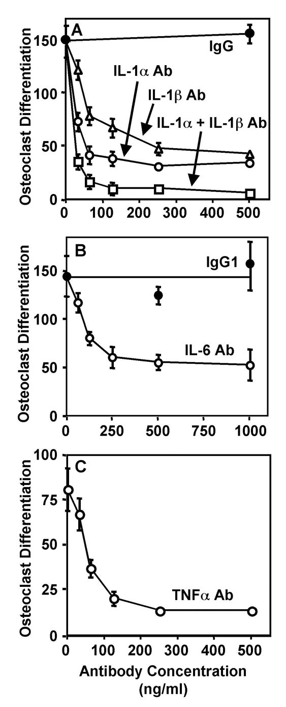
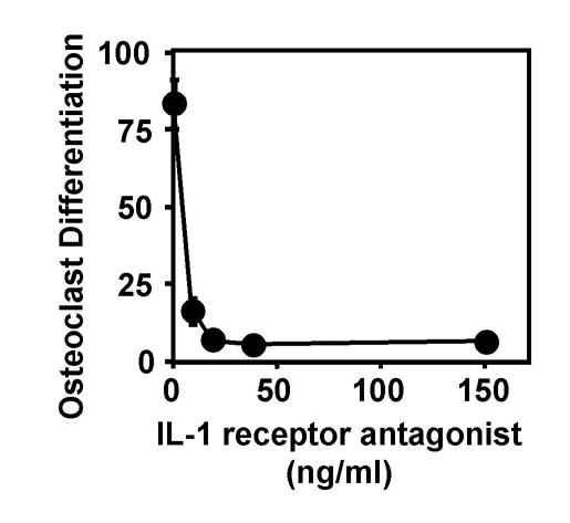
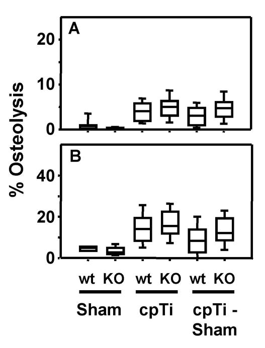
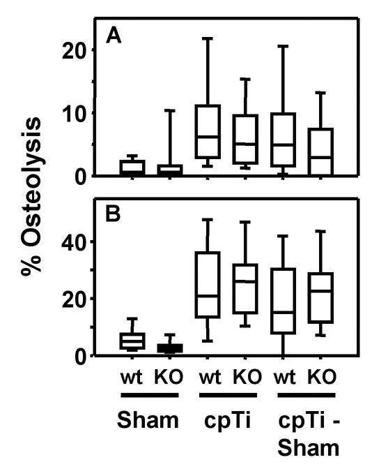
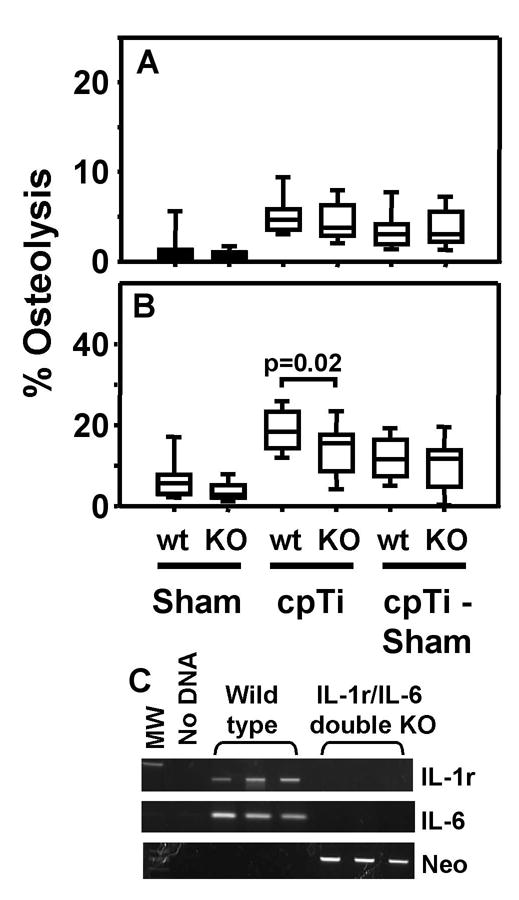
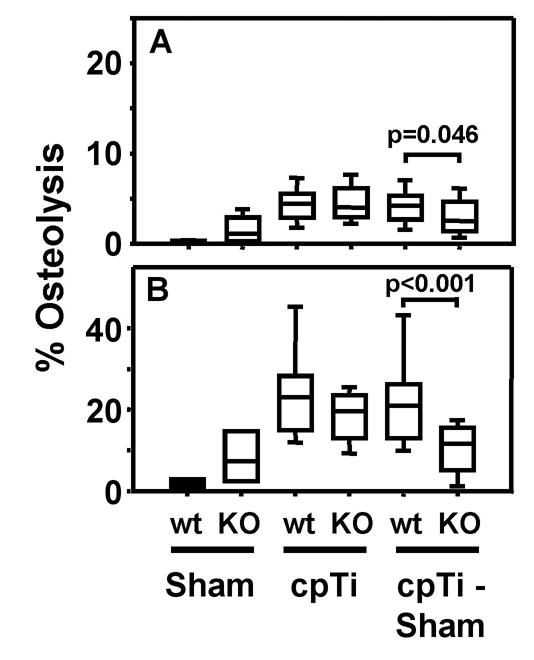
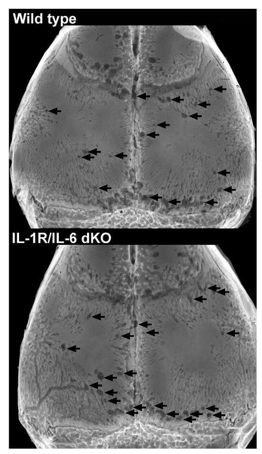
Pro-inflammatory cytokines, such as IL-1, IL-6, and TNF, are considered to be major mediators of osteolysis and ultimately aseptic loosening. This study demonstrated that synergistic interactions among these cytokines are required for the in vitro stimulation of osteoclast differentiation by titanium particles. In contrast, genetic knock out of these cytokines or their receptors does not protect murine calvaria from osteolysis induced by titanium particles. Thus, the extent of osteolysis was not substantially altered in single knock out mice lacking either the IL-1 receptor or IL-6. Osteolysis also was not substantially altered in double knock out mice lacking both the IL-1 receptor and IL-6 or in double knock out mice lacking both TNF receptor-1 and TNF receptor-2. The differences between the in vivo and the cell culture results make it difficult to conclude whether the pro-inflammatory cytokines contribute to aseptic loosening. One alternative is that in vivo experiments are more physiological and that therefore the current results do not support a role for the pro-inflammatory cytokines in aseptic loosening. We however favor the alternative that, in this case, the cell culture experiments can be more informative. We favor this alternative because the role of the pro-inflammatory cytokines may be obscured in vivo by compensation by other cytokines or by the low signal to noise ratio found in measurements of particle-induced osteolysis.


Figures

Figure 1
Figure 1
Neutralizing antibodies that target IL-1α (A), IL-1β (B), IL-6 (C), and TNFα (D) significantly inhibit osteoclast differentiation induced in vitro by titanium particles. Control cultures were incubated with non-immune goat IgG (A) or non-immune rat IgG1 (B). Osteoclast differentiation was assessed by measuring the number of TRAP-positive multinuclear cells/cm2. N = 5–6 for all groups.
Figure 2
Figure 2
Recombinant IL-1 receptor antagonist significantly inhibits osteoclast differentiation induced in vitro by titanium particles. Osteoclast differentiation was assessed by measuring the number of TRAP-positive multinuclear cells/cm2. N = 5–6 for all groups.
Figure 3
Figure 3
IL-1 receptor single knock out mice are not protected from titanium particle-induced osteolysis. Osteolysis was measured in the entire parietal bones (A) and in the suture regions (B). Data are presented as box plots showing the 10th, 25th, 50th, 75th, and 90th, percentiles. In A, n = 16–18 for first and second groups, n = 48 for the third and fifth groups, and n = 34 for the fourth and sixth groups. In B, n = 8–9 for the first and second groups, n = 24 for the third and fifth groups, and n = 17 for the fourth and sixth groups.
Figure 4
Figure 4
IL-6 single knock out mice are not protected from titanium particle-induced osteolysis. Osteolysis was measured in the entire parietal bones (A) and in the suture regions (B). Data are presented as box plots showing the 10th, 25th, 50th, 75th, and 90th, percentiles. In A, n = 22–28 for all groups. In B, n = 11–14 for all groups.
Figure 5
Figure 5
IL-1 receptor/IL-6 double knock out mice are not protected from titanium particle-induced osteolysis. Osteolysis was measured in the entire parietal bones (A) and in the suture regions (B). Data are presented as box plots showing the 10th, 25th, 50th, 75th, and 90th, percentiles. In A, n = 26–36 for all groups. In B, n = 13–18 for all groups. Panel C shows genotyping of the IL-1 receptor/IL-6 double knock out mice. PCR amplicons from three representative wild type and three representative double knock mice are shown. Tail clip DNA was amplified with primers specific for the IL-1 receptor gene, the IL-6 gene, and the neomycin cassette, respectively.
Figure 6
Figure 6
TNF receptor 1/TNF receptor 2 double knock out mice are not protected from titanium particle-induced osteolysis. Osteolysis was measured in the entire parietal bones (A) and in the suture regions (B). Data are presented as box plots showing the 10th, 25th, 50th, 75th, and 90th, percentiles. In A, n = 12 for the first and second groups and n = 24–26 for the third, fourth, fifth, and sixth groups. In B, n = 6 for the first and second groups and n = 12–13 for the third, fourth, fifth, and sixth groups.
Figure 7
Figure 7
Represenative examples of titanium-induced osteolysis in wild type and IL-1 receptor/IL-6 double knock out. The images are of the skull with the per cent osteolysis in the suture region that is closest to the median for each group. Arrows indicate some of the osteolytic areas.

Similar articles

Cited by

References

    1. Wright TM, Goodman SB, editors. Implant Wear in Total Joint Replacement. Rosemount, Ill: American Academy of Orthopaedic Surgeons; 2001.
    1. Greenfield EM, Bi Y, Ragab AA, Goldberg VM, Nalepka JL, Seabold JM. Does endotoxin contribute to aseptic loosening of orthopaedic implants? J Biomed Mater Res B Appl Biomater. 2005;72B:179–85. - PubMed
    1. Greenfield E. Particulate Matter and Host Reactions. In: Wnek G, Bowlin G, editors. Encyclopedia of Biomaterials and Biomedical Engineering. New York: Taylor Francis; 2006. - DOI
    1. Hicks DG, Judkins AR, Sickel JZ, Rosier RN, Puzas JE, O’Keefe RJ. Granular histiocytosis of pelvic lymph nodes following total hip arthroplasty. The presence of wear debris, cytokine production, and immunologically activated macrophages. J Bone Joint Surg. 1996;78A:482–96. - PubMed
    1. Xu J, Konttinen Y, Lassus J, Natah S, Ceponis A, Solovieva S, Aspenberg P, Santavirta S. Tumor necrosis factor-alpha (TNF-alpha) in loosening of total hip replacement (THR) Clin Exp Rheumatol. 1996;14:643–8. - PubMed

Publication types

MeSH terms