. 2007 Feb;14(2):207-18.
doi: 10.1016/j.acra.2006.10.016.

Initial clinical experience with microwave breast imaging in women with normal mammography

Affiliations

Initial clinical experience with microwave breast imaging in women with normal mammography

Paul M Meaney et al. Acad Radiol. 2007 Feb.

Abstract

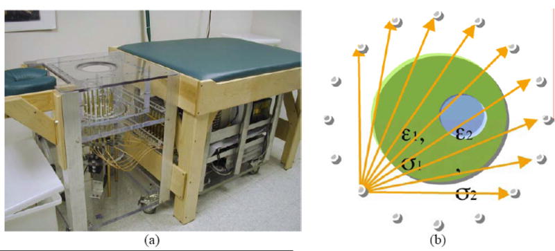
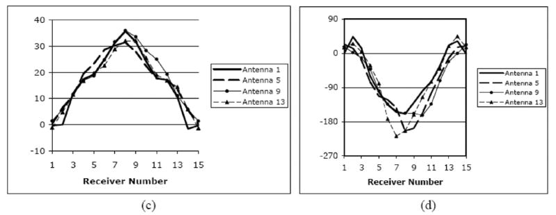
Rationale and objectives: We have developed a microwave tomography system for experimental breast imaging.

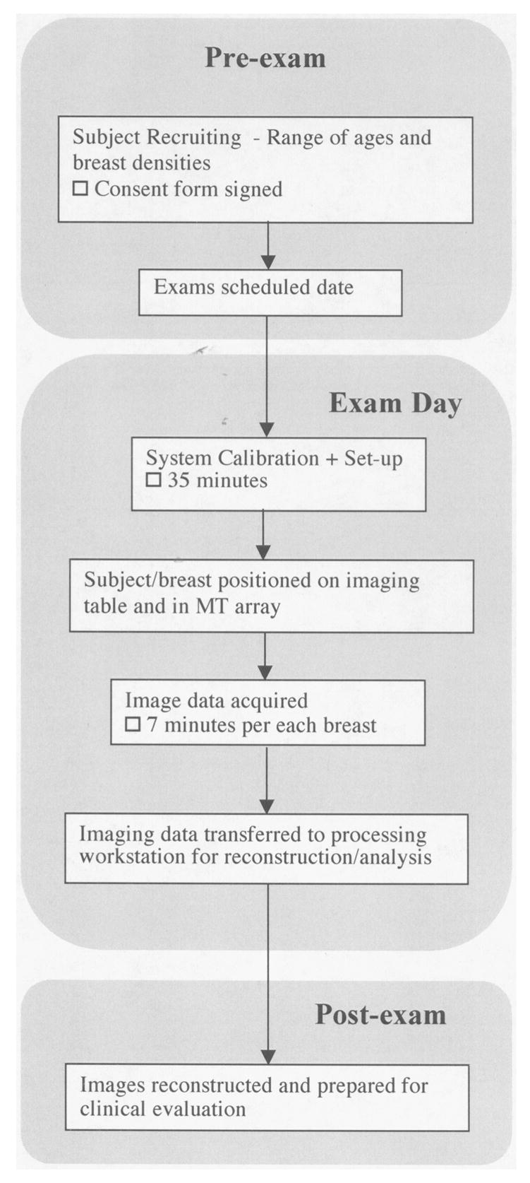
Materials and methods: In this article, we illustrate a strategy for optimizing the coupling liquid for the antenna array based on in vivo measurement data. We present representative phantom experiments to illustrate the imaging system's ability to recover accurate property distributions over the range of dielectric properties expected to be encountered clinically. To demonstrate clinical feasibility and assess the microwave properties of the normal breast in vivo, we summarize our initial experience with microwave breast exams of 43 women with negative mammography according to the Breast Imaging Reporting and Data System (BI-RADS 1).

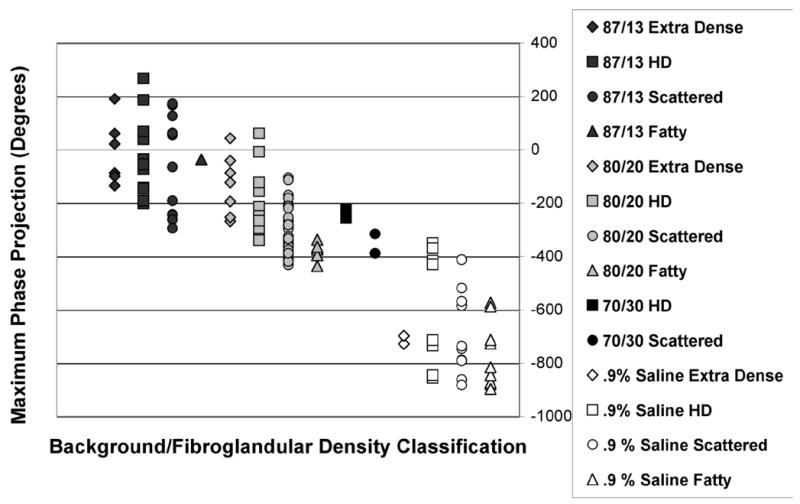
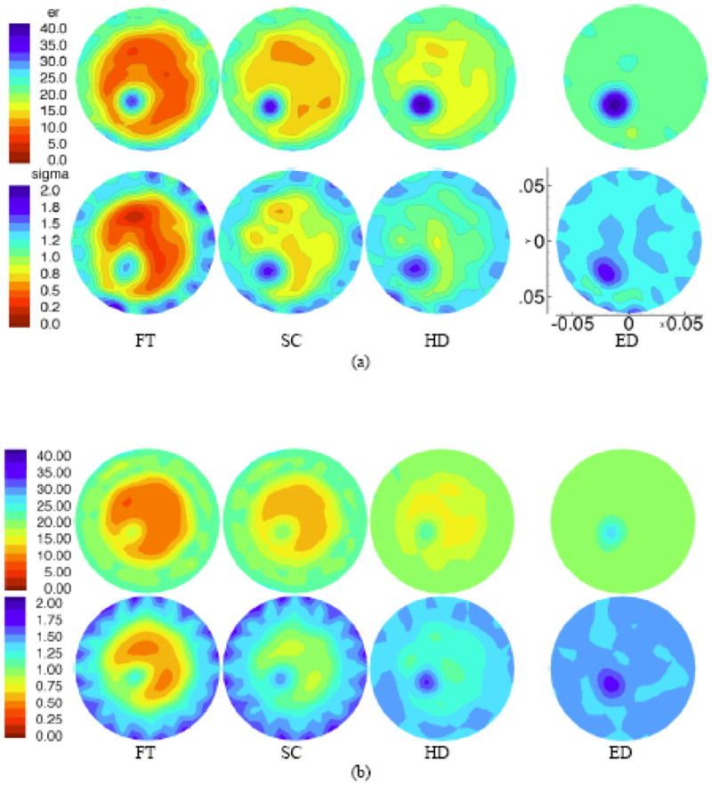
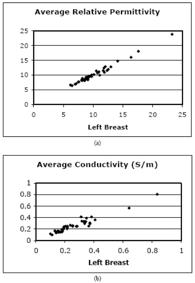
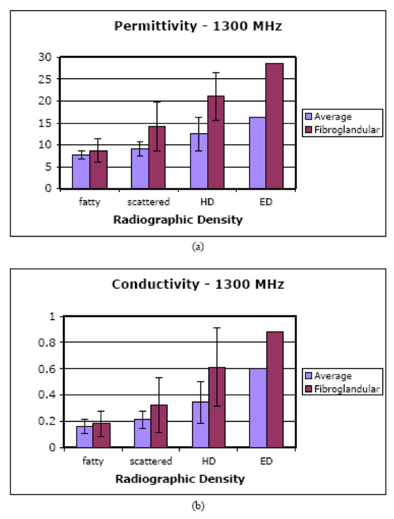
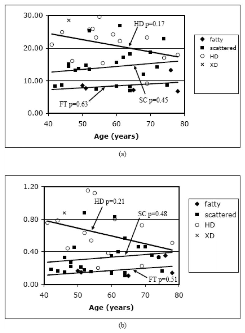
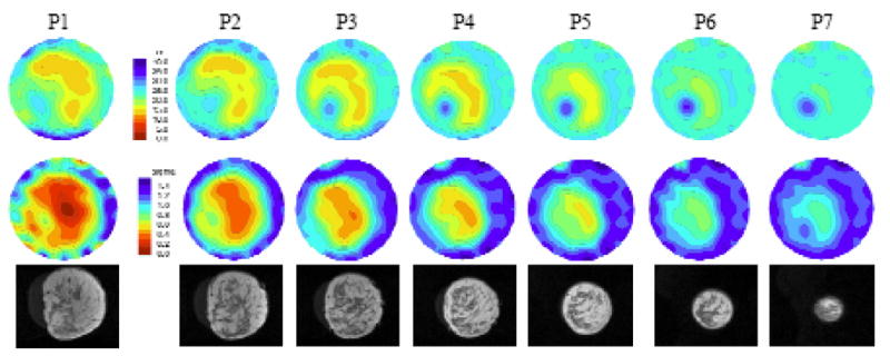
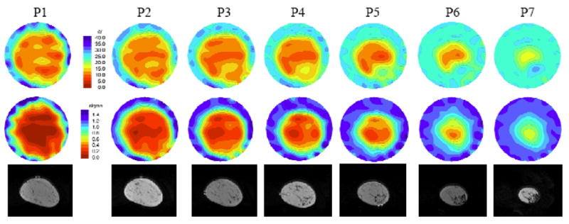
Results: The clinical results show a high degree of bilateral symmetry in the whole breast average microwave properties. Focal assessments of microwave properties are associated with breast tissue composition evaluated through radiographic density categorization verified through magnetic resonance image correlation in selected cases. Specifically, both whole-breast average and local microwave properties increase with increasing radiographic density, in which the latter exhibits a more substantial rise.

Conclusion: These findings support our hypothesis that water content variations in the breast play an influential role in dictating the overall dielectric property distributions and indicate that the microwave properties in the breast are more heterogeneous than previously believed based on ex vivo property measurements reported in the literature.


Figures

Figure 1
Figure 1
(a) Photograph of the clinical microwave breast imaging prototype showing the illumination tank, exam platform and electronics cart (underneath bed); (b) 2D schematic diagram of the illumination and reception configuration; and representative (c) amplitude and (d) phase projections for a set of measured clinical data for a single imaging plane at 1300 MHz (data for only 4 of the 16 illuminations are shown).
Figure 1
Figure 1
(a) Photograph of the clinical microwave breast imaging prototype showing the illumination tank, exam platform and electronics cart (underneath bed); (b) 2D schematic diagram of the illumination and reception configuration; and representative (c) amplitude and (d) phase projections for a set of measured clinical data for a single imaging plane at 1300 MHz (data for only 4 of the 16 illuminations are shown).
Figure 2
Figure 2
Typical phantom experiment with liquid containers suspended from above the tank and integrated with an alignment fixture for accurate positioning.
Figure 3
Figure 3
Flow graph of the process followed for patient recruiting, examination and data processing.
Figure 4
Figure 4
Maximum phase projection data from 900 MHz imaging experiments of 147 volunteers utilizing several different coupling baths (0.9 % saline, and 70:30, 80:20, and 87:13 glycerin:water coupling baths) for women with a range of breast densities.
Figure 5
Figure 5
1300 MHz permittivity (top) and conductivity (bottom) images for a (a) 10 cm phantom with a 2 cm inclusion, and a (b) 7.5 cm phantom with a 1 cm inclusion, respectively. Phantom designations (left to right): (FT) fatty, (SC) scattered, (HD) heterogeneously dense, and (ED) extremely dense have microwave properties which mimic those determined from in vivo images (see text for details). The imaging FOV is 13.5 cm in diameter in all cases and properties are reported on the common scales shown (left most image pair). Spatial dimensions are also the same in each case and a representative scale (in meters) is shown on the bottom right most image for the larger phantom case.
Figure 6
Figure 6
Scatter plots of the 1300 MHz average (a) permittivity and (b) conductivity for the right versus left breasts of the 43 normal subjects.
Figure 7
Figure 7
Bar graphs of the 1300 MHz overall average and fibroglandular average (a) permittivity and (b) conductivity values as a function of breast density for the 43 normal subjects.
Figure 8
Figure 8
Scatter plots of the 1300 MHz fibroglandular average (a) permittivity and (b) conductivity values grouped by radiographic density and graphed as a function of patient age. P-values for the trend lines are shown for each density category.
Figure 9
Figure 9
Microwave (top row – permittivity and middle row – conductivity at 1100 MHz) and coregistered MR (bottom row) images in the same anatomically coronal view for the right breast of a woman with heterogeneously dense tissue. The labels (P1 to P7) above each image correspond to the plane of acquisition relative to the chestwall. The property and spatial scales are the same for all images. The microwave image FOV is fixed at 13.5 cm in diameter.
Figure 10
Figure 10
Microwave (top row – permittivity and middle row – conductivity at 1100MHz) and coregistered MR (bottom row) images in the same anatomically coronal view for the left breast of a woman with fatty to scattered radiographic density. P1 though P7 (labels above each image column) indicate microwave tomograms spaced 1 cm apart beginning near the chestwall.

References

    1. American Cancer Society. Breast cancer facts and figures 2003–2004. American Cancer Society; Atlanta, GA: 2004.
    1. American College of Radiology (ACR) ACR Breast Imaging Reporting and Data System, Breast Imaging Atlas. 4. Reston, VA: American College of Radiology; 2003. ACR BI-RADS - Mammography.
    1. Benjamin R, Craddock IJ, Hilton GS, Litobarski S, McCutcheon E, Nilavalan R, Crisp GN. Microwave detection of buried mines using non-contact, synthetic near-field focusing. IEE Proceedings - Radar, Sonar and Navigation. 2001;148:233–240.
    1. Bolomey J-C, Izadnegahdar A, Jofre L, Pichot C, Peronnet G, Solaimani M. Microwave Diffraction Tomography for Biomedical Applications. IEEE Transactions on Microwave Theory and Techniques. 1982;82:1998 – 2000.
    1. Bolomey J-C, Jofre L, Peronnet G. On the Possible Use of Microwave-Active Imaging for Remote Thermal Sensing. IEEE Transactions on Microwave Theory and Techniques. 1983;83:777–781.