. 2007 Apr 1;92(7):2621-32.
doi: 10.1529/biophysj.106.100776. Epub 2007 Jan 19.

The sodium-coupled neutral amino acid transporter SNAT2 mediates an anion leak conductance that is differentially inhibited by transported substrates

Affiliations

The sodium-coupled neutral amino acid transporter SNAT2 mediates an anion leak conductance that is differentially inhibited by transported substrates

Zhou Zhang et al. Biophys J. .

Abstract

The sodium-coupled neutral amino acid transporter SNAT2 mediates cellular uptake of glutamine and other small, neutral amino acids. Here, we report the existence of a leak anion pathway associated with SNAT2. The leak anion conductance was increased by, but did not require the presence of, extracellular sodium. The transported substrates L-alanine, L-glutamine, and alpha-(methylamino)isobutyrate inhibited the anion leak conductance, each with different potency. A transporter with the mutation H-304A did not catalyze alanine transport but still catalyzed anion leak current, demonstrating that substrate transport is not required for anion current inhibition. Both the substrate and Na+ were able to bind to the SNAT2H-304A transporter normally. The selectivity sequence of the SNAT2H-304A anion conductance was SCN->>NO3->I->Br->Cl->Mes-. Anion flux mediated by the more hydrophobic anion SCN- was not saturable, whereas nitrate flux demonstrated saturation kinetics with an apparent Km of 29 mM. SNAT2, which belongs to the SLC38 family of transporters, has to be added to the growing number of secondary, Na+-coupled transporters catalyzing substrate-gated or leak anion conductances. Therefore, we can speculate that such anion-conducting pathways are general features of Na+-transporting systems.


Figures

FIGURE 1
FIGURE 1
Sequence alignment of the putative sixth transmembrane domain of the SLC38 (SNAT) family. The highly conserved SNAT2 His-304 residue mutated to alanine is highlighted.
FIGURE 2
FIGURE 2
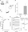
Functional characterization of SNAT2 expressed in HEK293 cells. (A) Currents induced by application of 10 mM alanine to control cells (left panel) and SNAT2-expressing cells (right panel). The bath solution contained 140 mM NaMes, and the pipette solution contained 140 mM KMes at 0 mV transmembrane potential. (B) Statistical analysis of transport currents between SNAT2WT-expressing and nontransfected cells (control). Leak currents were subtracted. The large error bar of the currents in the SNAT2-expressing cells is caused by the up to threefold differences in expression levels between different cells. (C) The apparent affinity for the substrate L-alanine of SNAT2WT was determined by recording substrate-induced transport current as a function of [alanine] at 0 mV. (D) Voltage dependence of representative L-alanine-induced transport currents. Saturating concentrations of L-alanine (10 mM) were applied in 140 mM NaMes at the time indicated by the bar. (E) Average alanine-induced current-voltage relationships in nontransfected cells (control, open circles) and SNAT2WT-expressing cells (solid squares).
FIGURE 3
FIGURE 3
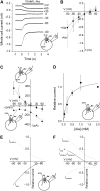
SNAT2 catalyzes an uncoupled leak anion conductance that is inhibited by alanine. (A) Voltage dependence of alanine-sensitive SNAT2WT currents in the presence of 140 mM intracellular KSCN and 140 mM extracellular NaMes. A saturating concentration of L-alanine (10 mM) was applied at t = 0 s, as indicated by the bar. (B) Current-voltage relationships in SNAT2WT-expressing cells in the absence (open squares) and in the presence (solid squares) of 10 mM L-alanine in the presence of 140 mM intracellular SCN. The dashed lines show currents in nontransfected control cells recorded under the same conditions (both dashed lines are superimposable). (C) Current-voltage relationships of L-alanine-sensitive currents (10 mM alanine) in SNAT2WT obtained after subtraction of the current in the absence from that in the presence of alanine with 140 mM SCN present only in the pipette (solid squares), and 140 mM SCN only present on the extracellular side (solid triangles). The open circles show data from control cells not expressing SNAT2 in the presence of intracellular SCN. (D) The apparent affinity for L-alanine of SNAT2WT was determined by recording the alanine-sensitive current as a function of [alanine] at 0 mV in the presence of 140 mM extracellular SCN. (E) Simulation of the leak anion current (Ileak) and the alanine-inhibition of the leak anion current (Iinhibition) obtained from Eq. 1. (F) Simulation of total (Itotal) L-alanine-sensitive currents. The total current in the presence of intracellular SCN (Itotal) was calculated as the sum of the alanine-inhibited leak anion current component (Iihibition = −Ileak) and the alanine-induced transport current component (Itransport).
FIGURE 4
FIGURE 4
The extent of inhibition of the leak anion conductance depends on the transported substrate. (A) Transport currents activated at 0 mV by application of L-glutamine (bottom panel) and MeAIB (top panel) to SNAT2 at concentrations indicated by the bar (Mes was used as the anion on both sides of the membrane). (B) Average current-voltage relationships of substrate-sensitive currents induced by 10 mM glutamine (solid squares) and 10 mM MeAIB (open circles) to SNAT2 in the presence of 140 mM intracellular SCN (n = 4). The main anion in the bath buffer was Mes. (C) Statistical analysis of reversal potentials for substrate-induced currents for alanine, glutamine, and MeAIB. The ionic conditions were as in (B).
FIGURE 5
FIGURE 5
Intracellular [SCN] determines the reversal potential of the alanine-sensitive current. (A) Mean current amplitudes for various internal [SCN] in the presence of 10 mM external L-alanine. SCN was substituted equimolarly with Mes. The currents were normalized to the currents recorded at +60 mV. (B) Reversal potential-[SCN] relationship of SNAT2WT anion currents.
FIGURE 6
FIGURE 6
SNAT2H-304A mediates the leak anion conductance but little alanine transport activity. (A) Comparison of alanine-induced transport currents between SNAT2H-304A-expressing and nontransfected cells (control). The bath solution contained NaMes, and the pipette solution contained KMes. Leak currents were subtracted. (B) Voltage dependence of SNAT2H-304A alanine-sensitive currents in the presence of 140 mM intracellular KSCN and 140 mM extracellular NaMes. Saturating concentrations of L-alanine (10 mM) were applied at t = 0 s, as indicated by the bar. (C) Current-voltage relationships of SNAT2H-304A currents in the absence (open squares) and in the presence (solid squares) of 10 mM L-alanine with intracellular 140 mM SCN. (D) Current-voltage relationship of L-alanine-sensitive currents in SNAT2H-304A (10 mM alanine) with SCN present only in the pipette (solid squares), SCN only present on the extracellular side (solid triangles). The open circles show data from control cells not expressing SNATH-304A in the presence of intracellular SCN.
FIGURE 7
FIGURE 7
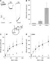
Leak anion currents are activated by extracellular Na+. Whole-cell current recordings were performed with a KSCN-based pipette solution (140 mM) and at 0 mV transmembrane potential. (A) Comparison of typical leak anion currents induced by the application of 140 mM extracellular Na+ between nontransfected cells (control), SNAT2WT, and SNAT2H-304A-expressing cells, as indicated by the gray bars. (B) Statistical analysis of average Na+-induced currents as the ones shown in (A). (C) and (D) show leak anion currents as a function of extracellular [Na+] for SNATWT (C) and SNAT2H-304A (D), respectively (solid circles, after subtracting the unspecific currents, open squares, determined from nontransfected cells). The solid lines represent fits to the Hill equation with a Hill coefficient of n = 1. The open circles in C and D are results from the original experiments before subtraction of the unspecific leak currents (open squares) determined in nontransfected control cells.
FIGURE 8
FIGURE 8
Leak anion currents are present in the absence of extracellular Na+. (A) Typical original data show the currents in response to a double solution-exchange protocol, first from NMGCl to NaCl, and second from NaCl to NaCl + 10 mM alanine, as indicated by the bars (top). The pipette contained 140 mM SCN. (B) Current-voltage relationships of the alanine-sensitive anion current in the absence of Na+ determined by subtraction from currents obtained at time points A-B, as indicated by the arrows in (A).
FIGURE 9
FIGURE 9
Anion selectivity of the transporter-associated anion conductance in SNAT2H-304A-expressing cells. (A) Responses after a solution exchange from 0 mM alanine to 10 mM alanine (indicated by the bar) at membrane potentials ranging from −90 to +60 mV. The pipette solution contained 10 mM KSCN and 130 mM KMes, the bath solution contained 126 mM NaSCN. (B) Voltage dependence of alanine-sensitive currents in an external solution containing 126 mM Mes (▴), Cl (∇), Br (▵), I (□), formula image (•), and SCN (▪) in the presence of 126 mM external Na+ with 10 mM KSCN plus 130 mM internal KMes; 10 mM L-alanine was applied.
FIGURE 10
FIGURE 10
Anion currents carried by formula image are saturable. (A) Typical currents induced by alanine (10 mM) at various external [SCN] concentrations at a holding potential of 0 mV. (B) and (C) show the determination of apparent Km values for SCN (B) and formula image (C) of anion binding to SNAT2H-304A by measuring the alanine-sensitive anion current at different extracellular [anion] (126 mM Na+). The solid lines represent fits to the Hill equation with a Hill coefficient of n = 1. For SCN no Km can be obtained because the current does not saturate within the concentration range tested. For formula image, the Km is 29 ± 8 mM.
FIGURE 11
FIGURE 11
Assignment of the anion conductance to specific states in the kinetic model of amino acid-Na+ cotransport by SNAT2. T and T′ are the transporters with the amino acid (aa) and Na+-binding sites exposed to the extracellular side and the cytoplasm, respectively. The anion-conducting states are indicated by the curved arrows with thicker arrows indicating higher anion conductance.

Similar articles

Cited by

References

    1. Johnson, L. W., and C. H. Smith. 1988. Neutral amino acid transport systems of microvillous membrane of human placenta. Am. J. Physiol. 254:C773–C780. - PubMed
    1. Sugawara, M., T. Nakanishi, Y. J. Fei, W. Huang, M. E. Ganapathy, F. H. Leibach, and V. Ganapathy. 2000. Cloning of an amino acid transporter with functional characteristics and tissue expression pattern identical to that of system A. J. Biol. Chem. 275:16473–16477. - PubMed
    1. Bode, B. P. 2001. Recent molecular advances in mammalian glutamine transport. J. Nutr. 131:2475S–2485S. - PubMed
    1. Yao, D., B. Mackenzie, H. Ming, H. Varoqui, H. Zhu, M. A. Hediger, and J. D. Erickson. 2000. A novel system A isoform mediating Na+/neutral amino acid cotransport. J. Biol. Chem. 275:22790–22797. - PubMed
    1. Shotwell, M. A., D. W. Jayme, M. S. Kilberg, and D. L. Oxender. 1981. Neutral amino acid transport systems in Chinese hamster ovary cells. J. Biol. Chem. 256:5422–5427. - PubMed

Publication types

LinkOut - more resources