. 2007 Jan;19(1):237-55.
doi: 10.1105/tpc.106.047720. Epub 2007 Jan 19.

The Arabidopsis irregular xylem8 mutant is deficient in glucuronoxylan and homogalacturonan, which are essential for secondary cell wall integrity

Affiliations

The Arabidopsis irregular xylem8 mutant is deficient in glucuronoxylan and homogalacturonan, which are essential for secondary cell wall integrity

Staffan Persson et al. Plant Cell. 2007 Jan.

Abstract

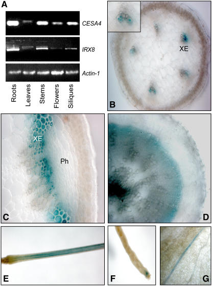
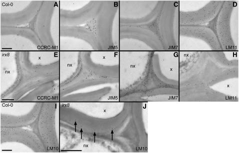
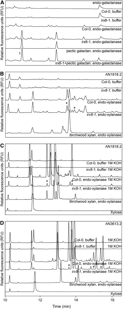
The secondary cell wall in higher plants consists mainly of cellulose, lignin, and xylan and is the major component of biomass in many species. The Arabidopsis thaliana irregular xylem8 (irx8) mutant is dwarfed and has a significant reduction in secondary cell wall thickness. IRX8 belongs to a subgroup of glycosyltransferase family 8 called the GAUT1-related gene family, whose members include GAUT1, a homogalacturonan galacturonosyltransferase, and GAUT12 (IRX8). Here, we use comparative cell wall analyses to show that the irx8 mutant contains significantly reduced levels of xylan and homogalacturonan. Immunohistochemical analyses confirmed that the level of xylan was significantly reduced in the mutant. Structural fingerprinting of the cell wall polymers further revealed that irx8 is deficient in glucuronoxylan. To explore the biological function of IRX8, we crossed irx8 with irx1 (affecting cellulose synthase 8). The homozygous irx1 irx8 exhibited severely dwarfed phenotypes, suggesting that IRX8 is essential for cell wall integrity during cellulose deficiency. Taken together, the data presented show that IRX8 affects the level of glucuronoxylan and homogalacturonan in higher plants and that IRX8 provides an important link between the xylan polymer and the secondary cell wall matrix and directly affects secondary cell wall integrity.


Figures

Figure 1.
Figure 1.
Phenotypic Features of irx8 Mutant Plants. (A) Relative positions of T-DNA insertions for the irx8 mutants. (B) to (E) Alterations in growth of irx8 mutant plants. (B) Seven-week-old plants showing shorter inflorescence stems in irx8 mutants (right) compared with the wild type (left). Bar = 5 cm. (C) Seven-week-old rosette leaves from irx8-1 (bottom) are significantly smaller compared with wild-type (top) leaves. (D) irx8 plants had smaller flowers with short anthers (arrow) and reduced amounts of pollen. The female organs appeared normal. (E) The siliques from irx8 mutants were significantly smaller and contained no seeds. (F) Hand-cut transverse sections from wild-type (left) and irx8-1 (right) stems stained with Toluidine blue. Arrows indicate collapsed xylem vessels. Ph, phloem; XE, xylem. (G) Transverse root sections (1-μm thick) from 7-week-old wild type (left) and irx8 (right) stained with Toluidine blue. Arrows indicate collapsed xylem vessels. Bars = 10 μm.
Figure 2.
Figure 2.
Early Onset of Irregular Xylem Phenotype in irx8. Hand-cut transverse sections from different developmental points of the stem for the wild type, irx8-1, and irx1 stained with Toluidine blue. Insets show enlarged xylem vessels. Arrows indicate collapsed xylem vessels. Bars = 20 μm. (A) to (C) Sections from base, middle, and top of wild-type stems, respectively. (D) to (F) Sections from base, middle, and top of irx8-1 stems, respectively. (G) to (I) Sections from base, middle, and top of irx1 stems, respectively.
Figure 3.
Figure 3.
Reduction in Cell Wall Thickness and Cell Size in irx8 Xylem Cells. Transmission electron microscopy of cell walls from the metaxylem region of wild-type and irx8-1 mature stems and roots. Bars = 2 μm. (A) Wild-type (left) and irx8-1 (right) stem sections. PW, residual primary cell walls. (B) Wild-type (left) and irx8-1 (right) root sections.
Figure 4.
Figure 4.
Expression of IRX8. (A) Expression of IRX8 and one of the secondary wall cellulose synthase subunits, CESA4, in different tissues assessed by RT-PCR. Actin was used as control. All tissues are from 7-week-old plants. (B) to (G) Expression patterns of the IRX8 gene assessed through an IRX8 promoter:GUS construct. The GUS expression is indicated by blue color. (B) Cross section of a 7-week-old stem at an expanding internode showing GUS staining at the developing metaxylem. Inset shows enlarged xylem vessels. XE, xylem vessels. (C) Cross section of a 7-week-old stem at the base showing GUS staining in both interfascicular fibers and xylem. Ph, phloem. (D) Cross section of 7-week-old root showing GUS staining associated with developing secondary xylem. (E) Part of inflorescence stem carrying siliques showing strong GUS expression. (F) Tip of silique. (G) Magnification of rosette leaf showing GUS staining associated with vascular strands.
Figure 5.
Figure 5.
Immunofluorescent Labeling of Transverse Sections of irx8 and Wild-Type Stems and Roots. Immunofluoscent labeling of 250-μm-thick transverse sections taken from root (rows 1 and 2) and stem (rows 3 and 4) tissues of 7-week-old Col-0 (rows 1 and 3) and irx8-1 (rows 2 and 4) plants. Antibodies used for labeling are indicated in the figure. Bars = 50 μm in (A) to (J) and 20 μm in (K) to (T). (A) to (E) Wild-type root sections labeled with CCRC-M1 (A), JIM5 (B), JIM7 (C), LM10 (D), and LM11 (E). (F) to (J) irx8 root sections labeled with CCRC-M1 (F), JIM5 (G), JIM7 (H), LM10 (I), and LM11 (J). (K) to (O) Wild-type stem sections labeled with CCRC-M1 (K), JIM5 (L), JIM7 (M), LM10 (N), and LM11 (O). (P) to (T) irx8 stem sections labeled with CCRC-M1 (P), JIM5 (Q), JIM7 (R), LM10 (S), and LM11 (T).
Figure 6.
Figure 6.
Immunogold Labeling of Xylem Cell Corners in 95-μm-Thick Transverse Sections Taken from Roots of 7-Week-Old Col-0 and irx8-1 Plants. (A) to (D) Wild-type sections labeled with CCRC-M1 (A), JIM5 (B), JIM7 (C), and LM11 (D). (E) to (H) irx8 sections labeled with CCRC-M1 (E), JIM5 (F), JIM7 (G), and LM11 (H). (I) Wild-type section labeled with LM10. (J) irx8 section labeled with LM10. The larger plate for irx8 LM10 labeling was included to show the sparse LM10 labeling (indicated by arrows). x, xylem cells; nx, nonxylem cells. Bars = 1 μm.
Figure 7.
Figure 7.
Capillary Electropherograms of Sugars and Oligosaccharides Released by Hydrolytic Enzyme Digests of Cell Walls from 7-Week-Old Stems. (A) Endogalactanase treatment. Commercial pectic galactan digested with endogalactanase was used as reference material. The 1 indicates galactose migration. RFU, relative fluorescence units. (B) Endoxylanase AN1818.2 (Bauer et al., 2006) treatment. Commercial birchwood xylan digested with endoxylanase AN1818.2 was used as reference material. (C) Endoxylanase AN1818.2 treatment of 1 M KOH fractions. (D) Endoxylanase AN3613.2 treatment of 1 M KOH fractions. The asterisk indicates peaks that differ between wild-type and irx8 mutants.
Figure 8.
Figure 8.
MALDI-TOF Mass Spectra from Xylanase-Treated Polysaccharides Present in 1 M KOH Fractions. (A) and (B) MALDI-TOF mass spectra of 1 M KOH fractions from the wild type (A) and irx8 (B) after treatment with endoxylanase AN1818.2. (C) and (D) MALDI-TOF mass spectra of 1 M KOH fractions from the wild type (C) and irx8 (D) after treatment with endoxylanase AN3613.2. Arrows indicate absent peaks in irx8 mutant spectra. Symbols indicate the oligosaccharide structure corresponding to the peaks. Closed squares, xylose; open circles, glucuronic acid; Me, methyl. All ions are observed as monosodium adducts [M+Na]+.
Figure 9.
Figure 9.
Analyses of irx8 irx9 Double Mutants. (A) Whole-plant morphology of wild-type, irx9-2, irx8-2, and irx8-2 irx9-2 plants. Bar = 5 cm. (B) Agarose gels showing wild-type, irx8-2, irx9-2, and irx8-2 irx9-2 genotypes as assessed by PCR. The irx8 genotyping (top panel) contains both genomic primers and insert primer. The two bottom panels are PCRs with genomic primers (top) and insert primer (bottom) for irx9, respectively. (C) Neutral sugar composition from 7-week-old stems of wild-type, irx8-2, irx9-2, and irx8-2 irx9-2 plants. The amounts of sugar are presented as mean values (n = 5) of micrograms of sugar/milligrams of dry weight ± se.
Figure 10.
Figure 10.
Analyses of irx1 irx8 Double Mutants. (A) Agarose gels showing wild-type, irx1, irx8-2, and irx8-2 irx1 genotypes as assessed by PCR. Top panel shows bands from the wild type and mutants after restriction digest with BpiI, which does not cut in irx1. Bottom panel shows irx8 genotypes after PCR. (B) Rosette leaves from 4-week-old wild-type, irx1, irx8-2, and irx8-2 irx1 plants. Bar = 5 cm (C) Whole-plant morphology of wild-type, irx1, irx8-2, and irx8-2 irx1 (arrow) plants. Inset shows magnified irx8-2 irx1 plant. Bars = 5 cm. (D) Hand-cut transverse sections from 7-week-old stems for wild-type and irx8-2 irx1 plants. Arrows indicate severely deformed xylem cells. XE, xylem; Ph, phloem. Bar = 50 μm. (E) irx8-2 irx1 mutant phenotype setting flowers and siliques directly on rosette. Bar = 5 cm.

Similar articles

Cited by

References

    1. Alonso, J., et al. (2003). Genome-wide insertional mutagenesis of Arabidopsis thaliana. Science 301 653–657. - PubMed
    1. Aspeborg, H., et al. (2005). Carbohydrate-active enzymes involved in the secondary cell wall biogenesis in hybrid aspen. Plant Physiol. 137 983–997. - PMC - PubMed
    1. Awano, T., Takabe, K., and Fujita, M. (2002). Xylan deposition on secondary wall of Fagus crenata fiber. Protoplasma 219 106–115. - PubMed
    1. Bauer, S., Vasu, P., Mort, A.J., and Somerville, C.R. (2005). Cloning, expression, and characterization of an oligoxyloglucan reducing end-specific xyloglucanobiohydrolase from Aspergillus nidulans. Carbohydr. Res. 340 2590–2597. - PubMed
    1. Bauer, S., Vasu, P., Persson, S., Mort, A.J., and Somerville, C.R. (2006). Development and application of a suite of polysaccharide degrading enzymes for analyzing plant cell walls. Proc. Natl. Acad. Sci. USA 103 11417–11422. - PMC - PubMed

Publication types

MeSH terms